

Localization of corin and atrial natriuretic peptide expression in human renal segments
- PMID: 27343265
- PMCID: PMC5237585
- DOI: 10.1042/CS20160398
Localization of corin and atrial natriuretic peptide expression in human renal segments
Abstract
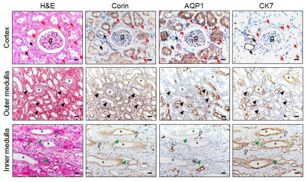
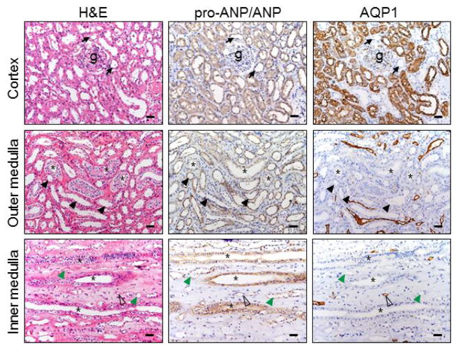
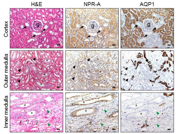
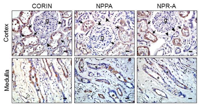
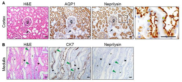
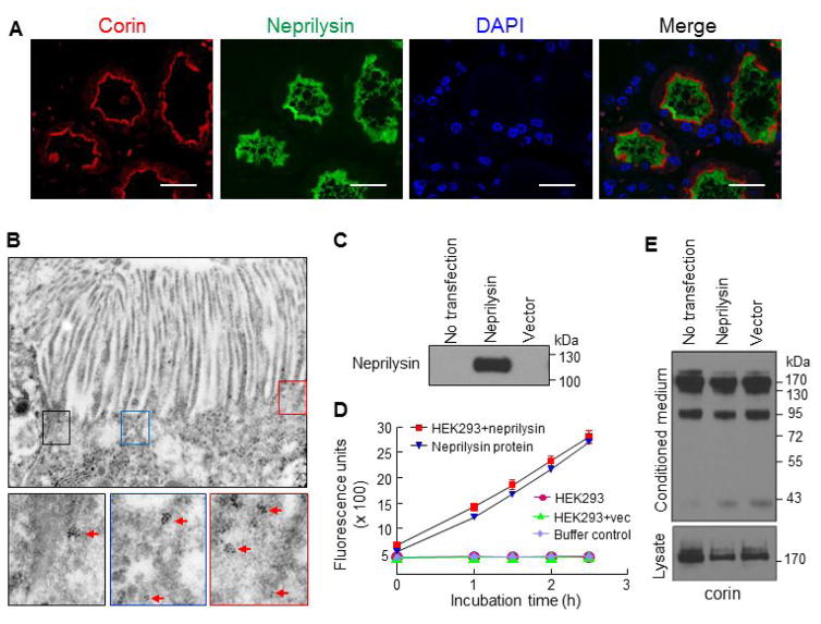
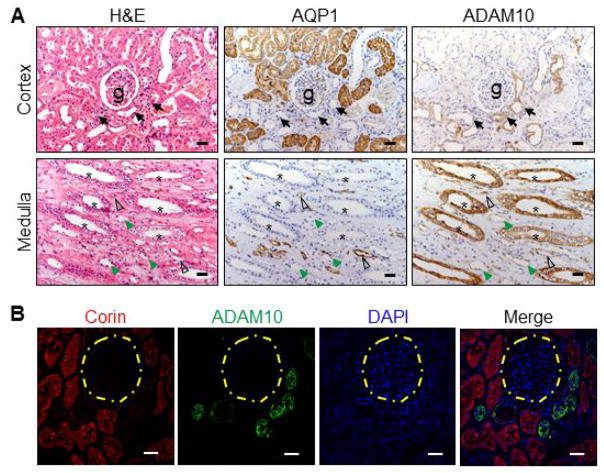
Atrial natriuretic peptide (ANP)-mediated natriuretic response is a well-established cardiac endocrine function. Corin is a transmembrane protease that activates ANP in the heart. Corin expression has been detected in non-cardiac tissues including the kidney. Here we examined corin, pro-ANP/ANP and natriuretic peptide receptor-A (NPR-A) expression in human renal segments. By immunostaining and in situ hybridization, we found similar corin, pro-ANP/ANP and NPR-A protein and mRNA expression in human renal segments. The expression was most abundant in the proximal convoluted tubules and the medullary connecting ducts. In the proximal tubules, corin protein was present in the apical membrane region underneath the brush border where the ANP-degrading protease neprilysin was abundant. These results suggest that corin-mediated pro-ANP activation may occur in renal segments and that locally produced ANP may act in an autocrine manner to regulate sodium and water reabsorption in situ Our results also point to the proximal convoluted tubules as a major site for local ANP action. Such a renal corin/ANP autocrine mechanism may differ from the cardiac corin/ANP endocrine mechanism in regulating sodium homoeostasis under physiological and pathological conditions.
Keywords: atrial natriuretic peptide; corin; natriuretic peptide receptor; neprilysin.
© 2016 The Author(s). published by Portland Press Limited on behalf of the Biochemical Society.
Conflict of interest statement
Declarations of interest None.
Figures
Similar articles
-
Role of corin in the regulation of blood pressure.Curr Opin Nephrol Hypertens. 2017 Mar;26(2):67-73. doi: 10.1097/MNH.0000000000000297. Curr Opin Nephrol Hypertens. 2017. PMID: 27898523 Free PMC article. Review.
-
Corin, a transmembrane cardiac serine protease, acts as a pro-atrial natriuretic peptide-converting enzyme.Proc Natl Acad Sci U S A. 2000 Jul 18;97(15):8525-9. doi: 10.1073/pnas.150149097. Proc Natl Acad Sci U S A. 2000. PMID: 10880574 Free PMC article.
-
Aberrant pro-atrial natriuretic peptide/corin/natriuretic peptide receptor signaling is present in maternal vascular endothelium in preeclampsia.Pregnancy Hypertens. 2018 Jan;11:1-6. doi: 10.1016/j.preghy.2017.12.001. Epub 2017 Dec 7. Pregnancy Hypertens. 2018. PMID: 29523263 Free PMC article.
-
Development of a novel, sensitive cell-based corin assay.Biochem Pharmacol. 2019 Feb;160:62-70. doi: 10.1016/j.bcp.2018.12.009. Epub 2018 Dec 13. Biochem Pharmacol. 2019. PMID: 30553787
-
The endocrine basis of the cardio-renal axis: New perspectives regarding corin.Physiol Rep. 2024 Jul;12(13):e16105. doi: 10.14814/phy2.16105. Physiol Rep. 2024. PMID: 38942727 Free PMC article. Review.
Cited by
-
Recombinant Soluble Corin Improves Cardiac Function in Mouse Models of Heart Failure.J Am Heart Assoc. 2021 Apr 6;10(7):e019961. doi: 10.1161/JAHA.120.019961. Epub 2021 Mar 24. J Am Heart Assoc. 2021. PMID: 33759549 Free PMC article.
-
Corin: A Key Mediator in Sodium Homeostasis, Vascular Remodeling, and Heart Failure.Biology (Basel). 2022 May 7;11(5):717. doi: 10.3390/biology11050717. Biology (Basel). 2022. PMID: 35625445 Free PMC article. Review.
-
Role of corin in the regulation of blood pressure.Curr Opin Nephrol Hypertens. 2017 Mar;26(2):67-73. doi: 10.1097/MNH.0000000000000297. Curr Opin Nephrol Hypertens. 2017. PMID: 27898523 Free PMC article. Review.
-
Atrial Natriuretic Peptide: Structure, Function, and Physiological Effects: A Narrative Review.Curr Cardiol Rev. 2021;17(6):e051121191003. doi: 10.2174/1573403X17666210202102210. Curr Cardiol Rev. 2021. PMID: 33530911 Free PMC article. Review.
-
Corin Is Downregulated in Renal Ischemia/Reperfusion Injury and Is Associated with Delayed Graft Function after Kidney Transplantation.Dis Markers. 2019 Jan 14;2019:9429323. doi: 10.1155/2019/9429323. eCollection 2019. Dis Markers. 2019. PMID: 30766618 Free PMC article.
References
-
- Rubattu S, Sciarretta S, Volpe M. Atrial natriuretic peptide gene variants and circulating levels: implications in cardiovascular diseases. Clin Sci (Lond) 2014;127:1–13. - PubMed
-
- Kuhn M. Molecular Physiology of Membrane Guanylyl Cyclase Receptors. Physiol Rev. 2016;96:751–804. - PubMed
-
- Kuhn M. Structure, regulation, and function of mammalian membrane guanylyl cyclase receptors, with a focus on guanylyl cyclase-A. Circ Res. 2003;93:700–9. - PubMed
-
- Potter LR, Abbey-Hosch S, Dickey DM. Natriuretic peptides, their receptors, and cyclic guanosine monophosphate-dependent signaling functions. Endocr Rev. 2006;27:47–72. - PubMed
Publication types
MeSH terms
Substances
Grants and funding
LinkOut - more resources
Full Text Sources
Other Literature Sources
Miscellaneous