. 2016 Sep:102:175-86.
doi: 10.1016/j.biomaterials.2016.06.031. Epub 2016 Jun 23.

Nanoparticle tumor localization, disruption of autophagosomal trafficking, and prolonged drug delivery improve survival in peritoneal mesothelioma

Affiliations

Nanoparticle tumor localization, disruption of autophagosomal trafficking, and prolonged drug delivery improve survival in peritoneal mesothelioma

Rong Liu et al. Biomaterials. 2016 Sep.

Abstract

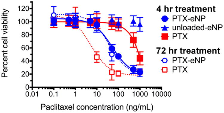
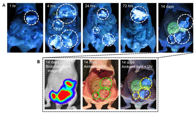
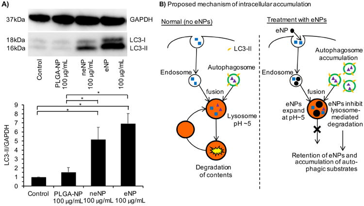
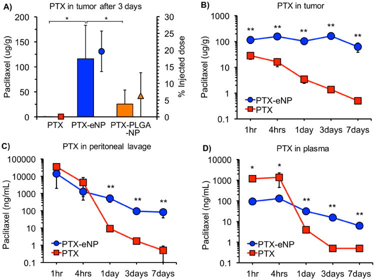
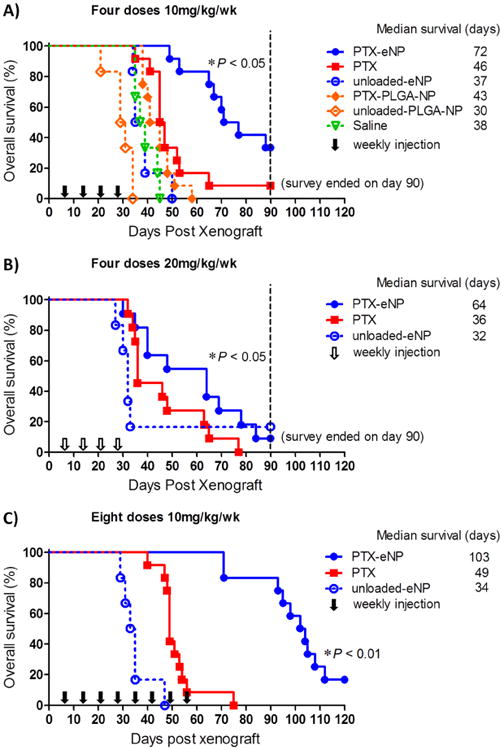
The treatment outcomes for malignant peritoneal mesothelioma are poor and associated with high co-morbidities due to suboptimal drug delivery. Thus, there is an unmet need for new approaches that concentrate drug at the tumor for a prolonged period of time yielding enhanced antitumor efficacy and improved metrics of treatment success. A paclitaxel-loaded pH-responsive expansile nanoparticle (PTX-eNP) system is described that addresses two unique challenges to improve the outcomes for peritoneal mesothelioma. First, following intraperitoneal administration, eNPs rapidly and specifically localize to tumors. The rate of eNP uptake by tumors is an order of magnitude faster than the rate of uptake in non-malignant cells; and, subsequent accumulation in autophagosomes and disruption of autophagosomal trafficking leads to prolonged intracellular retention of eNPs. The net effect of these combined mechanisms manifests as rapid localization to intraperitoneal tumors within 4 h of injection and persistent intratumoral retention for >14 days. Second, the high tumor-specificity of PTX-eNPs leads to delivery of greater than 100 times higher concentrations of drug in tumors compared to PTX alone and this is maintained for at least seven days following administration. As a result, overall survival of animals with established mesothelioma more than doubled when animals were treated with multiple doses of PTX-eNPs compared to equivalent dosing with PTX or non-responsive PTX-loaded nanoparticles.

Keywords: Autophagosome; Drug delivery; Mesothelioma; Nanoparticle; Paclitaxel; Tumor localization.


Conflict of interest statement

Disclosure of Conflicts of Interest: No potential conflicts of interest were disclosed.

Figures

Fig. 1
Fig. 1
Cytotoxicity of PTX-eNPs against human malignant mesothelioma cells using both short (4 hr; solid lines) and long/continuous (72 hr; dashed lines) duration exposure to treatments. MSTO-211H tumor cells were treated with PTX-eNPs or equivalent concentrations of PTX for the designated exposure time before cell viability was assessed via MTS assay. PTX-eNPs were significantly more potent after a 4 hr exposure than PTX (IC50 = 44.6 ng/mL vs 1,009 ng/mL, respectively). Unloaded-eNPs are not cytotoxic at any concentrations tested. Data are shown as mean ± SD (n ≥ 3).
Fig. 2
Fig. 2
Kinetics of cellular uptake of eNPs in malignant and non-malignant mesothelial cell lines. Cells were treated with Rho-eNPs for 0 (control), 2 or 8 hrs before washing and flow cytometric analysis. (A) Rho-eNP signal in human-derived MSTO-211H mesothelioma tumor cells increases rapidly (>98% cells compared to control) within 2 hrs of incubation (pink). (B) Rho-eNP uptake is slower (2% at 2 hrs (pink), 28% at 8 hrs (green)) in non-malignant human mesothelial LP-3 cells. Data are shown as mean ± SD (n ≥ 3).
Fig. 3
Fig. 3
Kinetics of PF-eNP localization to intraperitoneal mesothelioma tumors in vivo. The PF-eNPs emit bright white-blue light under UV-excitation. (A) Co-localization of PF-eNPs and tumors (white circles) in individual animals begins within 1-4 hrs after intraperitoneal PF-eNP injection and becomes more intense over several days. (B) Live bioluminescent (left), post-mortem ambient light (middle), and ambient/UV light combination (right) demonstrate co-localization of tumor burden (green circles) and PF-eNPs (yellow circles). Images are representative of three animals at each time point.
Fig. 4
Fig. 4
Quantification of LC3-II protein levels in eNP-treated cells by Western Blot. (A) Treatment of MSTO-211H tumor cells with PLGA-NPs does not significantly increase LC3-II levels compared to control. Treatment with eNPs and neNPs at 100 μg/mL significantly increases LC3-II levels, indicating the increased accumulation of autophagosomes. Results are normalized to the GAPDH protein level for each lane. Data are shown as mean ± SD (N = 5; * = P < 0.05). (B) Proposed mechanism by which eNPs (black filled circles) disrupt intracellular trafficking and lead to autophagosomal accumulation.
Fig. 5
Fig. 5
Paclitaxel concentrations in established tumor tissues, peritoneal lavage, and plasma after single bolus treatment injection given 14 days after inoculation with MSTO-211H tumor cells in NU/J mice. PTX-eNPs, PTX-PLGA-NPs or PTX (all at 10 mg/kg PTX dose) were injected intraperitoneally and tissues subsequently harvested for assessment of PTX levels. (A) PTX concentration in tumor tissues at 3 days (n ≥ 9). Columns represent tissue concentrations. Dots and bars represent percentage of injected dose in tumor tissues. (B, C, D) PTX concentrations in tumor, peritoneal lavage, and plasma as a function of time (n ≥ 3 per time point). Data are shown as mean ± SD (* P < 0.05, ** P < 0.001) Note: Tumor concentration data for PTX-eNPs in panel A was previously reported in [45] and is used for comparison. No other data from panels A, B, C, or D has been published.
Fig. 6
Fig. 6
Impact of multi-dose PTX-eNP regimen on animal survival in a murine model of established intraperitoneal mesothelioma. (A) Four weekly IP doses of PTX-eNPs nearly doubles overall survival. Weekly intraperitoneal treatments (arrows) were initiated 7 days after 5 million MSTO-211H/luc cells were injected. Animals received 10 mg/kg/dose PTX in PTX-eNPs, PTX-PLGA-NPs, or PTX, or a matched control treatment of unloaded-eNPs, unloaded-PLGA-NPs, or saline (*P < 0.05). (B) Impact of doubling PTX dose on overall survival. Animals received 4 doses of 20 mg/kg PTX as PTX-eNPs or PTX, or unloaded-eNPs (* P < 0.05). (C) Impact of doubling the number of doses of PTX on overall survival. Animals received eight-doses of 10 mg/kg PTX in PTX-eNPs or PTX, or an unloaded-eNP control (* P < 0.01).

References

    1. Chahinian AP, Pajak TF, Holland JF, Norton L, Ambinder RM, Mandel EM. Diffuse malignant mesothelioma. Prospective evaluation of 69 patients. Ann Intern Med. 1982;96:746–55. - PubMed
    1. Law MR, Gregor A, Hodson ME, Bloom HJ, Turner-Warwick M. Malignant mesothelioma of the pleura: a study of 52 treated and 64 untreated patients. Thorax. 1984;39:255–9. - PMC - PubMed
    1. Martini N, McCormack PM, Bains MS, Kaiser LR, Burt ME, Hilaris BS. Pleural mesothelioma. The Annals of thoracic surgery. 1987;43:113–20. - PubMed
    1. Garcia-Carbonero R, Paz-Ares L. Systemic chemotherapy in the management of malignant peritoneal mesothelioma. Eur J Surg Oncol. 2006;32:676–81. - PubMed
    1. Yan TD, Welch L, Black D, Sugarbaker PH. A systematic review on the efficacy of cytoreductive surgery combined with perioperative intraperitoneal chemotherapy for diffuse malignancy peritoneal mesothelioma. Ann Oncol. 2007;18:827–34. - PubMed

Publication types

MeSH terms