Comparative Study
. 2016 Sep:102:220-30.
doi: 10.1016/j.biomaterials.2016.06.025. Epub 2016 Jun 16.

Comparative biology of decellularized lung matrix: Implications of species mismatch in regenerative medicine

Affiliations
Comparative Study

Comparative biology of decellularized lung matrix: Implications of species mismatch in regenerative medicine

Jenna L Balestrini et al. Biomaterials. 2016 Sep.

Abstract

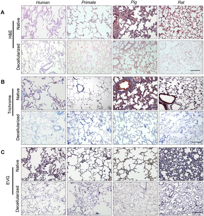
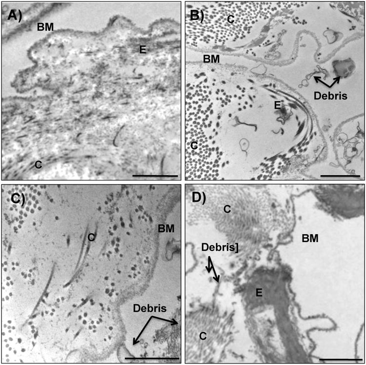
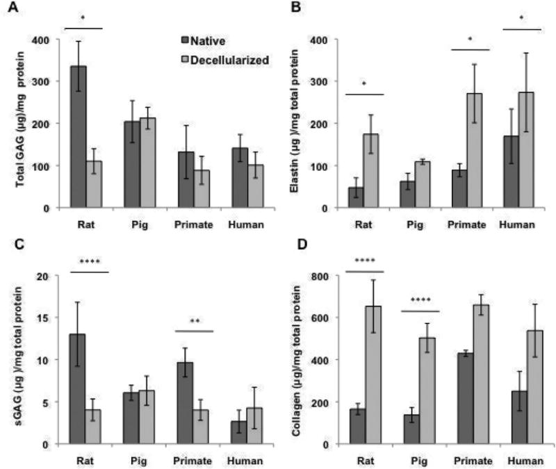
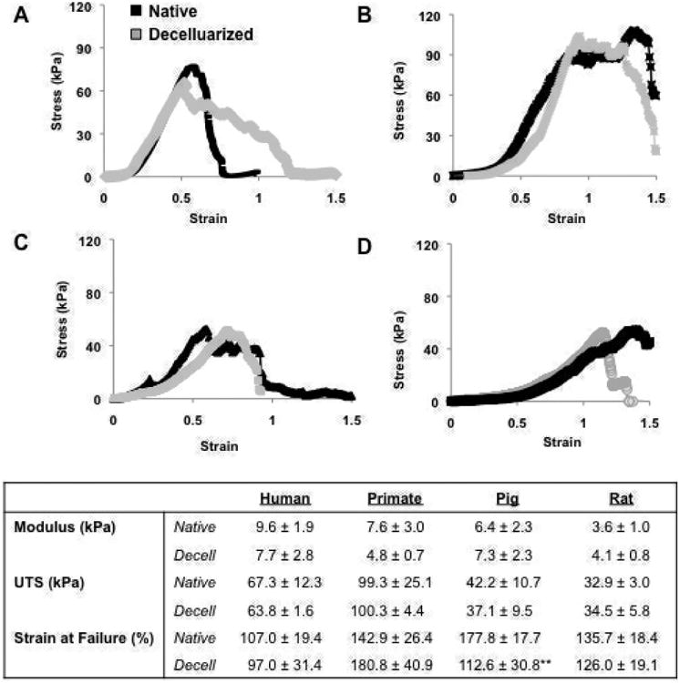
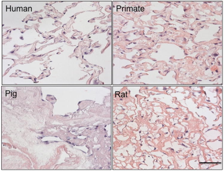
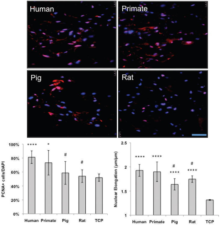
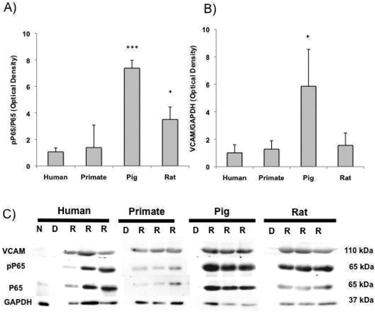
Lung engineering is a promising technology, relying on re-seeding of either human or xenographic decellularized matrices with patient-derived pulmonary cells. Little is known about the species-specificity of decellularization in various models of lung regeneration, or if species dependent cell-matrix interactions exist within these systems. Therefore decellularized scaffolds were produced from rat, pig, primate and human lungs, and assessed by measuring residual DNA, mechanical properties, and key matrix proteins (collagen, elastin, glycosaminoglycans). To study intrinsic matrix biologic cues, human endothelial cells were seeded onto acellular slices and analyzed for markers of cell health and inflammation. Despite similar levels of collagen after decellularization, human and primate lungs were stiffer, contained more elastin, and retained fewer glycosaminoglycans than pig or rat lung scaffolds. Human endothelial cells seeded onto human and primate lung tissue demonstrated less expression of vascular cell adhesion molecule and activation of nuclear factor-κB compared to those seeded onto rodent or porcine tissue. Adhesion of endothelial cells was markedly enhanced on human and primate tissues. Our work suggests that species-dependent biologic cues intrinsic to lung extracellular matrix could have profound effects on attempts at lung regeneration.

Keywords: Bioactivity; Decellularization; Extracellular matrix; Lung tissue engineering.


Figures

Fig. 1
Fig. 1. Characterization of human, primate, pig and rat decellularized lung matrix
(A) Representative native and decellularized lung tissue, as visualized by hematoxylin and eosin (H&E). Sections indicate maintenance of tissue architecture, removal of debris and blood, and lack of visible nuclear material. B) Preservation of collagen (blue) is visualized using Masson's Trichrome, and C) elastin (blue-black) by Verhoeff-Van Gieson staining. Scale bar = 100 μm applies to all panels.
Figure 2
Figure 2. TEM images of decellularized tissue
Representative transmission electron micrographs of A) human, B) primate, C) pig and D) rat decellularized tissue. Irrespective of species, decellularized lungs appeared to lack intact cellular bodies and retain basement membranes and characteristic normal architecture. Of the species tested, human decellularized tissue demonstrated the greatest homogeneous clearing of cellular debris. Collagen content appeared in clusters throughout the entirety of the primate lungs, and dispersed more sporadically throughout rat, pig and human tissue. Elastin did not appear highly concentrated in porcine tissue. Basement membrane (BM), collagen (C), elastin (E), and cellular debris (debris) are indicated with black arrows. Scale bar = 1 μm.
Figure 3
Figure 3. Matrix analyses of rat, porcine, primate and human decellularized lung tissue
Quantification of A) Total GAGs, B) elastin content, C) sGAGs, and D) collagen content of native and decellularized indicate the retention or loss of matrix post decellularization. For all ECM analysis, 3-5 pieces per lung were utilized and averaged (n=3, 3, 4, and 2 for rat, porcine, primate and human native and decellularized tissue). Quantitative values all normalized to dry starting tissue weight. * indicates p≤ 0.05, **p≤ 0.01, ***p≤ 0.001, and ****p≤ 0.0001.
Fig 4
Fig 4. Mechanical analysis of decellularized matrix
Representative stress strain curves of A) human, B) primate, C) pig, and D) rat native and decellularized tissue. B) Modulus, UTS, and Strain at Failure for native and decellularized lung tissue. ** indicates significance from native tissue at p ≤ 0.01.
Fig. 5
Fig. 5. Endothelial cell engraftment is species dependent
Human endothelial cells were seeded onto 1 cm2 tissue slices at a concentration of 1×10⁶ cells/slice for 3 days. Cells homogenously attached in both the primate and human tissue, heterogeneously throughout the rat tissue, and sparsely through the pig tissue. Scale bar = 100 μm applies to all panels.
Fig. 6
Fig. 6. Human endothelial cells demonstrate enhanced proliferation on human and primate matrix
Representative IF images of reseeded decellularized human, primate, pig and rat tissue. Human endothelial (VeraVec) cells were seeded onto 1 cm2 tissue slices at a concentration of 1,000,000 cells/slice for 3 days and stained for DAPI (in blue) PCNA (in red). The percentage positive cells (PCNA/DAPI) are noted in the bottom right hand of the images (n=4, average of 10 images). All cells on tissue were significantly more elongated than cells on TCP, yet only cells on human and primate tissues proliferated more than on TCP. Human cells proliferated significantly higher and were more elongated on human and primate tissue than on pig and rat tissues. Scale bar = 100 μm applies to all panels. * indicates p≤ 0.05, **p≤ 0.01, ***p≤ 0.001, and ****p≤ 0.0001 relative to TCP, # indicates p≤ 0.05 relative to human tissues.
Figure 7
Figure 7. Scaffold species source dictates EC inflammatory response
A) pP65/P65 and B) VCAM/GAPDH presence ECs seeded onto human, primate, rat and pig tissue. C) Representative western blots of native ‘N’, decellularized ‘D’ and recellularized ‘R’ rat, pig, primate and human lung slices immunoblotted for phosphorolated P65 (relative to P65) and VCAM/GAPDH (n≥3). * indicates p≤ 0.05, *** indicates p≤ 0.001 relative to ECs on human tissue.

References

    1. Yusen RD, Edwards LB, Kucheryavaya AY, Benden C, Dipchand AI, Dobbels F, Goldfarb SB, Levvey BJ, Lund LH, Meiser B, Stehlik J International Society for H, Lung T. The registry of the International Society for Heart and Lung Transplantation: thirty-first adult lung and heart-lung transplant report--2014; focus theme: retransplantation. J Heart Lung Transplant. 2014;33:1009–1024. - PubMed
    1. Ott HC, Clippinger B, Conrad C, Schuetz C, Pomerantseva I, Ikonomou L, Kotton D, Vacanti JP. Regeneration and orthotopic transplantation of a bioartificial lung. Nat Med. 2010;16:927–933. - PubMed
    1. Price AP, England KA, Matson AM, Blazar BR, Panoskaltsis-Mortari A. Development of a Decellularized Lung Bioreactor System for Bioengineering the Lung: The Matrix Reloaded. Tissue Eng Part A. 2010;16:2581–2591. - PMC - PubMed
    1. Nichols JE, Niles J, Riddle M, Vargas G, Schilagard T, Ma L, Edward K, La Francesca S, Sakamoto J, Vega S, Ogadegbe M, Mlcak R, Deyo D, Woodson L, McQuitty C, Lick S, Beckles D, Melo E, Cortiella J. Production and assessment of decellularized pig and human lung scaffolds. Tissue Eng Part A. 2013;19:2045–2062. - PMC - PubMed
    1. Bonvillain RW, Danchuk S, Sullivan DE, Betancourt AM, Semon JA, Eagle ME, Mayeux JP, Gregory AN, Wang G, Townley IK, Borg ZD, Weiss DJ, Bunnell BA. A nonhuman primate model of lung regeneration: detergent-mediated decellularization and initial in vitro recellularization with mesenchymal stem cells. Tissue Eng Part A. 2012;18:2437–2452. - PMC - PubMed

Publication types