. 2016 Jun 27:6:28862.
doi: 10.1038/srep28862.

Calcitonin attenuates cartilage degeneration and nociception in an experimental rat model of osteoarthritis: role of TGF-β in chondrocytes

Affiliations

Calcitonin attenuates cartilage degeneration and nociception in an experimental rat model of osteoarthritis: role of TGF-β in chondrocytes

Zhi-Hong Wen et al. Sci Rep. .

Abstract

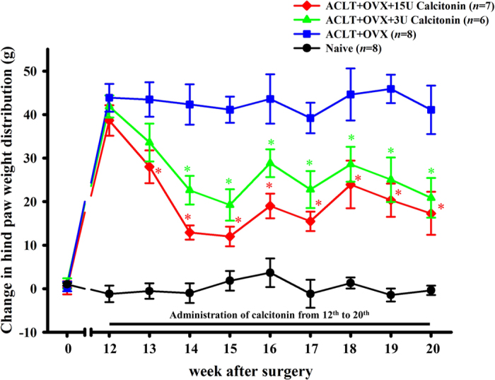
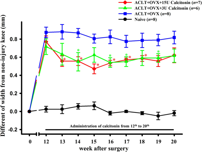
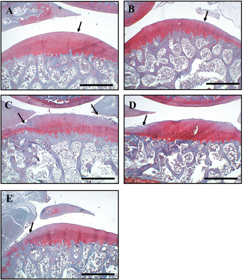
We investigated the role of the calcitonin (Miacalcin) in the progression of osteoarthritis (OA) and in nociceptive behavior in an experimental rat model of OA and osteoporosis. OA was induced by anterior cruciate ligament transection (ACLT) of the right knee and by bilateral ovariectomy (OVX) in Wistar rats. Nociceptive behaviors (secondary mechanical allodynia and weight-bearing distribution of the hind paws) were analyzed prior to surgery and every week, beginning at 12 weeks after surgery, up to 20 weeks. At 20 weeks, histopathological studies were performed on the cartilage of the knee joints. Immunohistochemical analysis was performed to examine the effect of calcitonin on transforming growth factor (TGF)-β1 expression in articular cartilage chondrocytes. Rats subjected to ACLT + OVX surgery showed obvious OA changes in the joints. Animals subjected to ACLT + OVX and treated with calcitonin showed significantly less cartilage degeneration and improved nociceptive tests compared with animals subjected to ACLT + OVX surgeries alone. Moreover, calcitonin increased TGF-β1 expression in chondrocytes in ACLT + OVX-affected cartilage. Subcutaneous injection of calcitonin (1) attenuated the development of OA, (2) concomitantly reduced nociception, and (3) modulated chondrocyte metabolism, possibly by increasing cellular TGF-β1 expression.


Figures

Figure 1
Figure 1. Time course of the anti-allodynic effect of calcitonin in ACLT-induced OA/OVX-induced osteoporosis nociception pain model.
Data are the mean ± SEM of the ipsilateral hind paw of each group. *P < 0.05 vs the ACLT + OVX group.
Figure 2
Figure 2. Time course of the effect of calcitonin on weight bearing of the contralateral (left) and ipsilateral (right) ACLT hind paws for 20 weeks after ACLT/OVX surgery.
The baseline was set as the weight-bearing differential prior to surgery. Data are the mean ± SEM of each group. *P < 0.05 vs the ACLT + OVX group.
Figure 3
Figure 3. Time course of the effects of calcitonin on bilateral hind knee joint widths after ACLT/OVX.
The data are the mean ± SEM of knee width, with baseline set as the widths prior to surgery. *P < 0.05 vs the ACLT + OVX group.
Figure 4
Figure 4. Histopathological evaluation of articular cartilage of the knee joint.
In both the naïve (A) and calcitonin 15 U (B) groups, the surface of the superficial cartilaginous layer was smooth (arrow) and the cartilage matrix was prominently stained with H/E. Specimens from the ACLT + OVX group (C) showed decreased cartilage thickness, disappearance of the surface layer cells (arrow), a fissure extending into the transitional and radial zones, and chondrocyte hypocellularity in the transitional and radial zones. The specimens from the ACLT + OVX + 3 U calcitonin group (D) and ACLT + OVX + 15 U calcitonin groups (E) showed mild irregularity of the surface layer, fibrillation of and fissures within the superficial cartilaginous layer (arrow), and slight diffuse hypercellularity in the transitional and radial zones. Stain, H/E; original magnification, ×40. Scale bar = 200 μm.
Figure 5
Figure 5. Distribution of TGF-β1 protein immunoreactivity and confocal double-immunofluorescent staining of TGF-β1 and articular chondrocyte marker doublecortin in the cartilage.
Positive immunoreactivity of the TGF-β1 protein is indicated by the red-brown color (arrows). The panels show the distribution of anti-TGF-β1 immunoreactivity in the cartilage of the (A) naïve, (B) calcitonin 15 U, (C) ACLT + OVX, (D) ACLT + OVX + 3 U calcitonin, and (E) ACLT + OVX + 15 U calcitonin groups. All samples were stained with antibodies against TGF-β1 protein. (F) Quantitative analysis showed that calcitonin significantly enhanced the number of TGF-β1-positive cells in the cartilage of ACLT + OVX knees. Scale bar = 100 μm. *P < 0.05 compared with the naïve group, #P < 0.05 compared with the ACLT + OVX group. Representative confocal immunofluorescence microscopy images showing the localization of TGF-β1 (G; green in color) and doublecortin (H; red in color) of articular cartilage in the ACLT + OVX + 15 U calcitonin group. Colocalization is indicated by yellow (I) and arrows. The confocal results showed that TGF-β1 was primarily co-localized with articular chondrocytes. Scale bars are 50 μm for all images.

References

    1. Pelletier J. P., Martel-Pelletier J. & Abramson S. B. Osteoarthritis, an inflammatory disease: potential implication for the selection of new therapeutic targets. Arthritis Rheum 44, 1237–1247, doi: 10.1002/1529-0131(200106) (2001). - DOI - PubMed
    1. Bajaj P., Baja P., Graven-Nielsen T. & Arendt-Nielsen L. Osteoarthritis and its association with muscle hyperalgesia: an experimental controlled study. Pain 93, 107–114, doi: 10.1016/S0304-3959(01)00300-1 (2001). - DOI - PubMed
    1. Lohmander L. S. et al.. Intra-articular hyaluronan injections in the treatment of osteoarthritis of the knee: a randomized, double blind, placebo controlled multicentre trial. Ann Rheum Dis 55, 424–431 (1996). - PMC - PubMed
    1. Høegh-Andersen P. et al.. Ovariectomized rats as a model of postmenopausal osteoarthritis: validation and application. Arthritis Res Ther 6, R169–180, doi: 10.1186/ar1152 (2004). - DOI - PMC - PubMed
    1. Sondergaard B. C., Ostergaard S., Christiansen C. & Karsdal M. A. The effect of oral calcitonin on cartilage turnover and surface erosion in the ovariectomized rat model. Arthritis Rheum 56, 2674–2678, doi: 10.1002/art.22797 (2007). - DOI - PubMed

Publication types