. 2016 Jun 27:6:28402.
doi: 10.1038/srep28402.

Involvement of Cl(-)/HCO3(-) exchanger SLC26A3 and SLC26A6 in preimplantation embryo cleavage

Affiliations

Involvement of Cl(-)/HCO3(-) exchanger SLC26A3 and SLC26A6 in preimplantation embryo cleavage

Yong Chao Lu et al. Sci Rep. .

Abstract

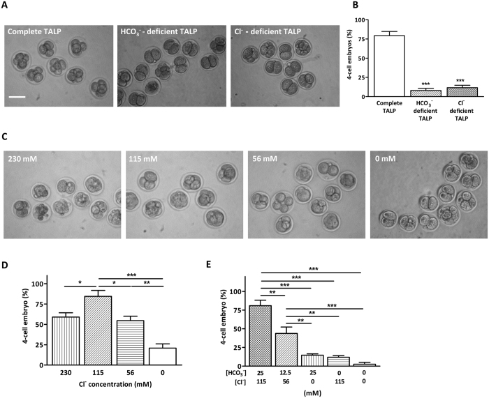
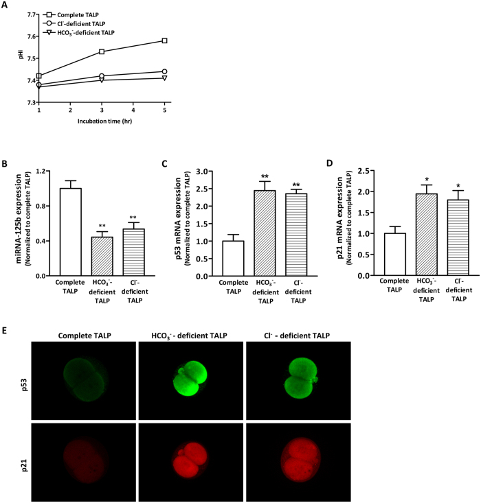
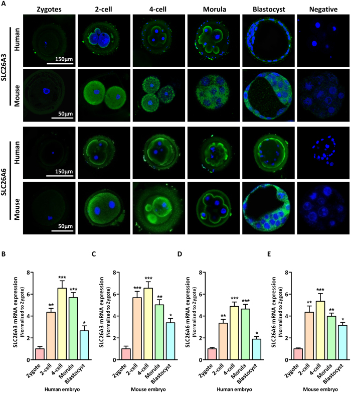
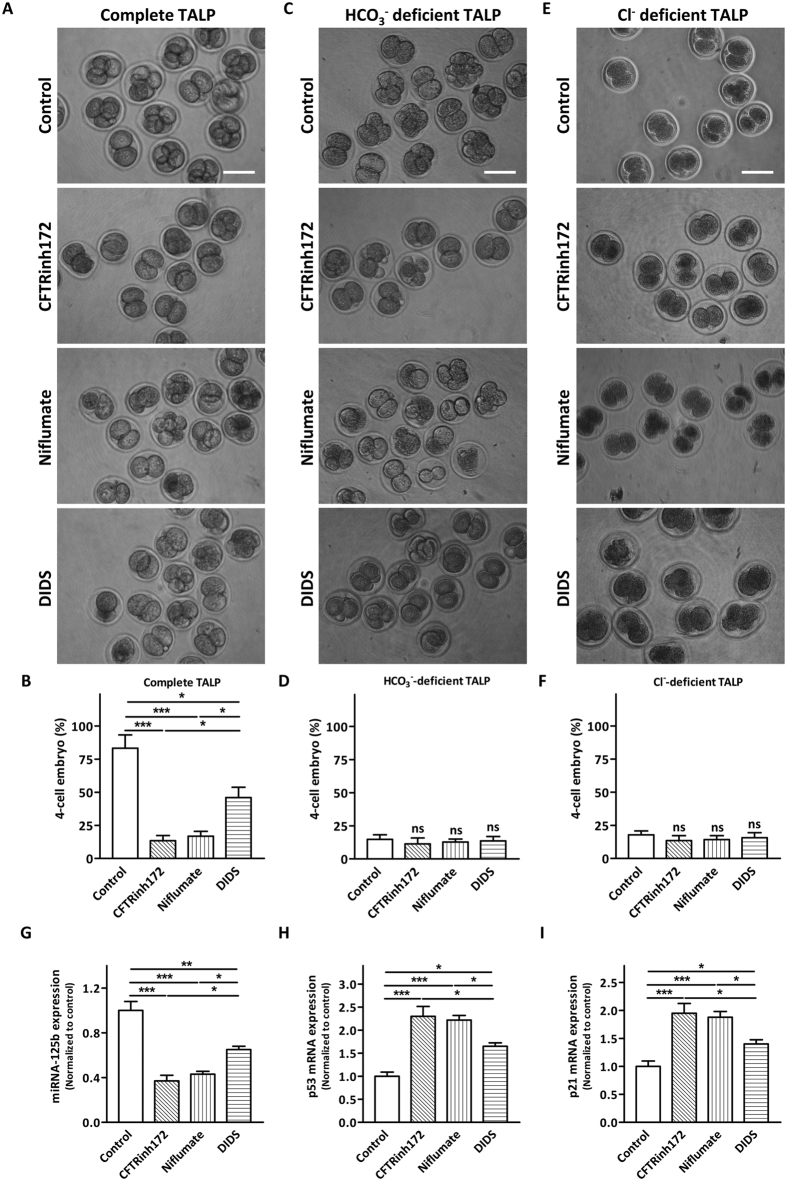
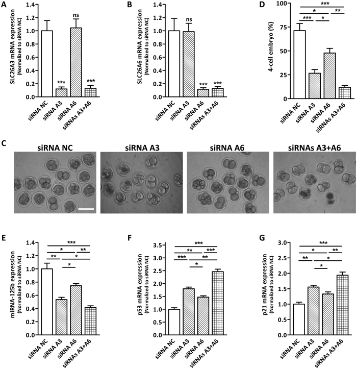
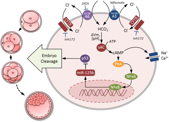
Bicarbonate (HCO3(-)) is essential for preimplantation embryo development. However, the mechanism underlying the HCO3(-) transport into the embryo remains elusive. In the present study, we examined the possible involvement of Cl(-)/HCO3(-) exchanger in mediating HCO3(-) transport into the embryo. Our results showed that depletion of extracellular Cl(-), even in the presence of HCO3(-), suppressed embryo cleavage in a concentration-dependent manner. Cleavage-associated HCO3(-)-dependent events, including increase of intracellular pH, upregulation of miR-125b and downregulation of p53, also required Cl(-). We further showed that Cl(-)/HCO3(-) exchanger solute carrier family 26 (SLC26) A3 and A6 were expressed at 2-cell through blastocyst stage. Blocking individual exchanger's activity by inhibitors or gene knockdown differentially decreased embryo cleavage and inhibited HCO3(-)-dependent events, while inhibiting/knocking down both produced an additive effect to an extent similar to that observed when CFTR was inhibited. These results indicate the involvement of SLC26A3 and A6 in transporting HCO3(-) essential for embryo cleavage, possibly working in concert with CFTR through a Cl(-) recycling pathway. The present study sheds light into our understanding of molecular mechanisms regulating embryo cleavage by the female reproductive tract.


Figures

Figure 1
Figure 1. Cl and HCO3- are both required for embryo cleavage.
(A) Four-cell embryo formation after 12 hours of 2-cell embryo culture in complete, Cl deficient or HCO3 deficient TALP medium (complete TALP: 25/32 embryos; HCO3 deficient TALP: 4/43 embryos; Cl deficient TALP: 7/39 embryos). Scale bar: 100 μm. (B) Summary of the results from A. *** indicates P < 0.001 (by one-way ANOVA, n = 4) when compared with control. (C) Four-cell embryo formation after 12 hours of 2-cell embryo culture in TALP medium with varying concentrations of Cl and constant HCO3 concentration (25 mM) (230 mM: 20/35 embryos; 115 mM: 29/33 embryos; 56 mM: 20/37 embryos; 0 mM: 7/30 embryos). Scale bar: 100 μm. (D) Summary of the results from C. * indicates P < 0.05, ** indicates P < 0.01, *** indicates P < 0.001 (by one-way ANOVA, n = 4). (E) Summary of the synergistic effect of HCO3 and Cl on embryo cleavage (0 mM HCO3 and 0 mM Cl: 1/40 embryos; 12.5 mM HCO3 and 56 mM Cl: 18/41 embryos; 0 mM HCO3 and 115 mM Cl: 5/42 embryos; 25 mM HCO3 and 0 mM Cl: 6/41 embryos; 25 mM HCO3 and 115 mM Cl: 30/37 embryos). ** indicates P < 0.01, *** indicates P < 0.001 (by one-way ANOVA, n = 4).
Figure 2
Figure 2. Involvement of Cl in HCO3-dependent signaling essential for embryo cleavage.
(A) Intracellular pH measurement of 2-cell embryos incubated in Cl-deficient, HCO3-deficient or complete TALP medium. Five micromolar BCECF was loaded for 30 min before determination of the pHi. The time course changes in pHi were shown (10 embryos/group). The experiments were repeated 4 times. (B–D) Quantitative real-time PCR result showing the expression of miRNA-125b (B), p53 (C) and p21 (D) in embryos incubated in HCO3-free or Cl-free condition compared to that incubated in complete TALP medium. ** indicates P < 0.01 (by one-way ANOVA, ~100 embryos in each experiment, n = 4). (E) Representative immunofluorescence results showing increased expression of p53 (green) and p21 (red) in 0 mM HCO3 condition or 0 mM Cl condition (5–10 embryos/group). Scale bar, 150 μm.
Figure 3
Figure 3. Expression of SLC26A3 and SLC26A6 in preimplantation embryos.
(A) Immunofluorescent results showing the localization of SLC26A3 (upper panel, green) and SLC26A6 (lower panel, green) protein in different stages of human and mouse preimplantation embryos. Nuclei were counterstained with DAPI (blue). (B–E) Quantitative real-time PCR results showing the levels of SLC26A3 (B,D) and SLC26A6 (C,E) mRNA expression during human (B,C) and mouse (D,E) preimplantation embryo development. * indicates P < 0.05, ** indicates P < 0.01, *** indicates P < 0.001 (by one-way ANOVA, ~100 embryos in each experiment, n = 4).
Figure 4
Figure 4. Effects of SLC26A3 and SLC26A6 inhibitors on embryo cleavage and HCO3-dependent signalling cascade.
(A–F) Effects of inhibiting CFTR (CFTRinh172, 10 μM), SLC26A3 (niflumate, 20 μM) and SLC26A6 (DIDS, 20 μM) on preimplantation embryo cleavage in complete TALP (A,B) control: 32/38 embryos; CFTRinh172: 6/34 embryos; niflumate: 9/35 embryos; DIDS: 14/40 embryos), HCO3-deficient TALP (C,D) control: 5/30 embryos; CFTRinh172: 7/31 embryos; niflumate: 4/32 embryos; DIDS: 6/31 embryos) and Cl-deficient TALP (E,F, control: 6/32 embryos; CFTRinh172: 4/31 embryos; niflumate: 5/30 embryos; DIDS: 6/33 embryos). Scale bar: 100 μm. Summary of the results are shown on the right panel (B,D,F). ns indicates P > 0.05, * indicates P < 0.05, ** indicates P < 0.01, *** indicates P < 0.001 (by one-way ANOVA, n = 4). (G–I) Quantitative real-time PCR showing the expression of miRNA-125b (G), p53 (H) and p21 (I) after CFTRinh172, niflumate or DIDS treatment. * indicates P < 0.05, ** indicates P < 0.01 (by one-way ANOVA, ~100 embryos in each experiment, n = 4).
Figure 5
Figure 5. Effect of SLC26A3 and/or SLC26A6 knockdown on embryo cleavage and HCO3-dependent signalling cascade.
(A,B) Quantitative real-time PCR result showing the expression of SLC26A3 (A) and SLC26A6 (B) mRNA in embryos after control siRNA (siRNA NC), SLC26A3 siRNA, SLC26A6 siRNA or both SLC26A3 and SLC26A6 siRNA injection at the 2-cell stage. *** indicates P < 0.001 (by one-way ANOVA, n = 4). (C) Four-cell embryo formation after 12 hours of embryo culture following siRNA NC, SLC26A3 siRNA, SLC26A6 siRNA or both SLC26A3 and SLC26A6 siRNA injection at 2-cell stage (siRNA NC: 32/45 embryos; SLC26A3 siRNA: 11/44 embryos; SLC26A6 siRNA: 21/40 embryos; SLC26A3 + A6 siRNA: 5/42 embryos). Scale bar: 100 μm. (D) Summary of the results from C. * indicates P < 0.05, ** indicates P < 0.01, *** indicates P < 0.001 (by one-way ANOVA, n = 4). (E–G) Quantitative real-time PCR showing the expression of miRNA-125b (E), p53 (F) and p21 (G) after siRNA NC, SLC26A6 siRNA, SLC26A3 siRNA or both SLC26A3 and SLC26A6 siRNA injection. * indicates P < 0.05, ** indicates P < 0.01, *** indicates P < 0.001 (by one-way ANOVA, ~100 embryos in each experiment, n = 4).
Figure 6
Figure 6. SLC26A3 and SLC26A6 work in concert with CFTR in regulating HCO3 transport in preimplantation embryo.
Working model for the regulation of early embryo cleavage by SLC26A3 and SLC26A6 in CFTR/HCO3-dependent activation of miR-125b. The HCO3 influx is mediated by SLC26A3 and SLC26A6 with an exchange of 2Cl/ HCO3. Apart from its reported role in conducting HCO3 directly, CFTR act as a Cl channel to provide a recycling pathway for Cl that is required for SLC26A3 and SLC26A6 function. The sites of action for inhibitors CFTRinh172, niflumate, and DIDS, as well as the intracellular HCO3-dependent events, are also shown. HCO3 influx mediated by the cooperative action of SLC26A3, SLC26A6 and CFTR activates soluble adenylyl cyclase (sAC) which increase the level of cAMP. This in turn activates PKA/NFkB signaling cascade which increases the expression of miR125b. Expression of miR125b is required for embryo cleavage through suppressing the expression of p53 and p21. Vm, membrane potential; [pH]i, intracellular pH.

Similar articles

Cited by

References

    1. Lu Y. C. et al. CFTR mediates bicarbonate-dependent activation of miR-125b in preimplantation embryo development. Cell research 22, 1453–1466, 10.1038/cr.2012.88 (2012). - DOI - PMC - PubMed
    1. Lu Y. C. et al. Small-conductance calcium-activated K(+) channels 3 (SK3) regulate blastocyst hatching by control of intracellular calcium concentration. Hum Reprod 27, 1421–1430, 10.1093/humrep/des060 (2012). - DOI - PubMed
    1. Salilew-Wondim D., Schellander K., Hoelker M. & Tesfaye D. Oviductal, endometrial and embryonic gene expression patterns as molecular clues for pregnancy establishment. Animal reproduction science 134, 9–18, 10.1016/j.anireprosci.2012.08.006 (2012). - DOI - PubMed
    1. Yoshinaga K. Progesterone and its downstream molecules as blastocyst implantation essential factors. American journal of reproductive immunology 72, 117–128, 10.1111/aji.12253 (2014). - DOI - PubMed
    1. Salehnia M. & Zavareh S. The effects of progesterone on oocyte maturation and embryo development. International journal of fertility & sterility 7, 74–81 (2013). - PMC - PubMed

Publication types