. 2016 Sep:75:36-43.
doi: 10.1016/j.mcn.2016.06.003. Epub 2016 Jun 21.

MicroRNA-1-associated effects of neuron-specific brain-derived neurotrophic factor gene deletion in dorsal root ganglia

Affiliations

MicroRNA-1-associated effects of neuron-specific brain-derived neurotrophic factor gene deletion in dorsal root ganglia

Elena Neumann et al. Mol Cell Neurosci. 2016 Sep.

Abstract

Background: MicroRNAs (miRNAs) regulate gene expression in physiological as well as in pathological processes, including chronic pain. Whether deletion of a gene can affect expression of the miRNAs that associate with the deleted gene mRNA remains elusive. We investigated the effects of brain-derived neurotrophic factor (Bdnf) gene deletion on the expression of miR-1 in dorsal root ganglion (DRG) neurons and its pain-associated downstream targets heat shock protein 60 (Hsp60) and connexin 43 (Cx43) in tamoxifen-inducible conditional knockout mice, Bdnf(fl/fl); Advillin-CreER(T2) (Bdnf cKO).

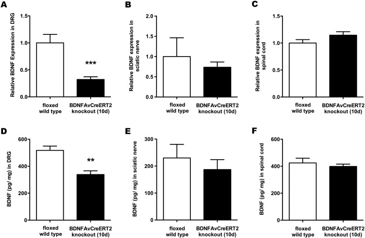
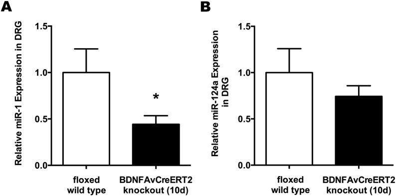
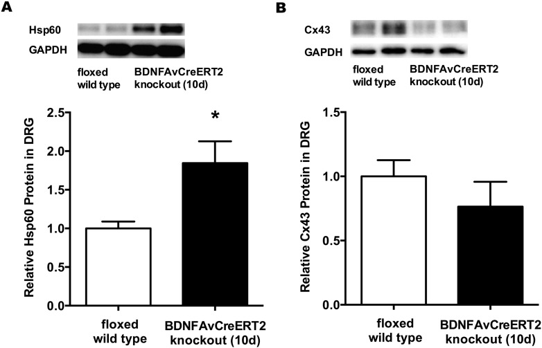
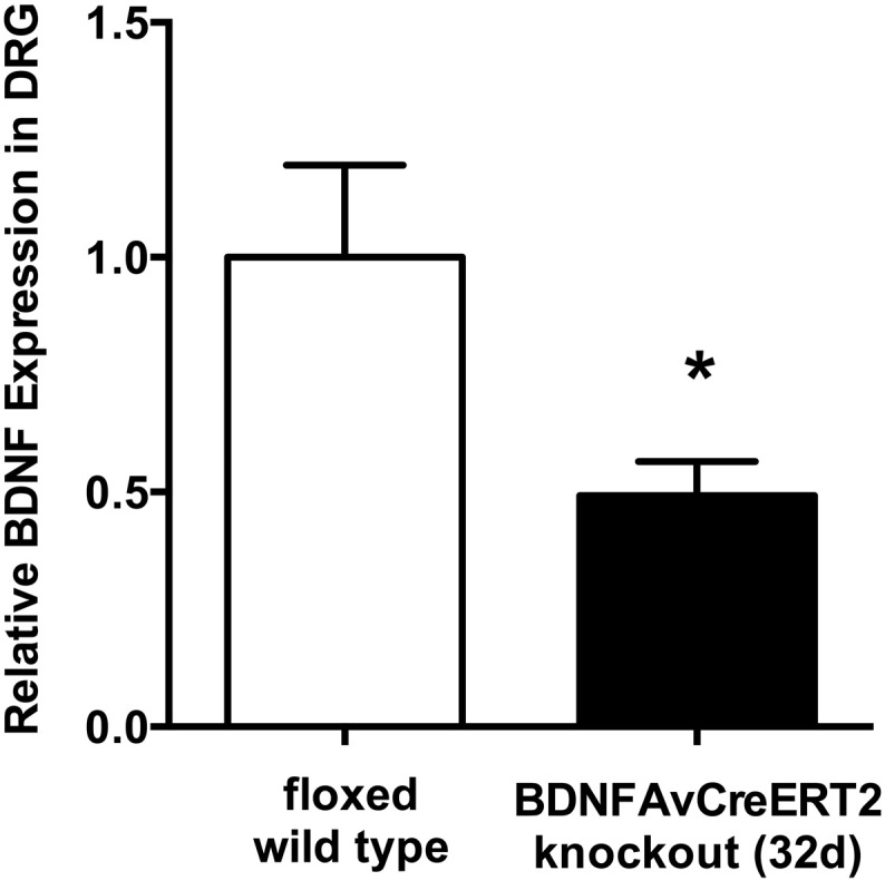
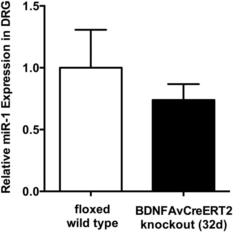
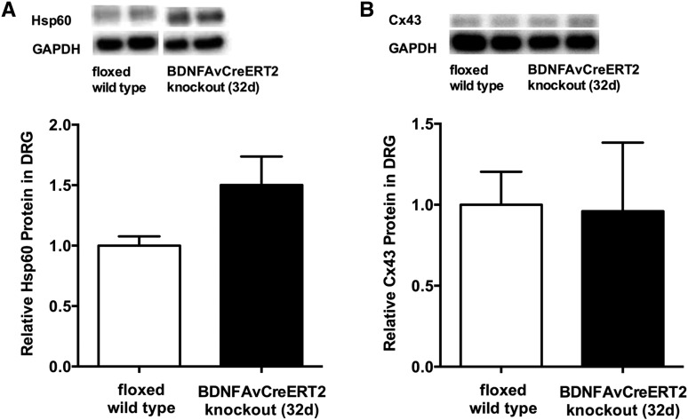
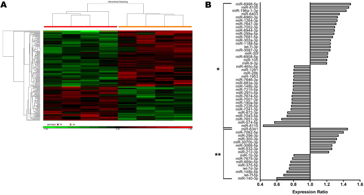
Results: Efficient Bdnf gene deletion was confirmed in DRG of Bdnf cKO mice by Real-Time qRT-PCR and ELISA 10days after completed tamoxifen treatment. In DRG, miR-1 expression was reduced 0.44-fold (p<0.05; Real-time qRT-PCR) in Bdnf cKO compared to floxed wildtype littermate control Bdnf(fl/fl) mice (WT). While Hsp60 protein expression was increased 1.85-fold (p<0.05; Western blot analysis), expression levels of Cx43 and the miR-1-associated transcription factors MEF2a and SRF remained unchanged. When analyzing Bdnf cKO mice 32days after complete tamoxifen treatment to investigate whether observed expression alterations remain permanently, we found no significant differences between Bdnf cKO and WT mice. However, miRNA microarray analysis revealed that 167 miRNAs altered (p<0.05) in DRG of these mice following Bdnf gene deletion.

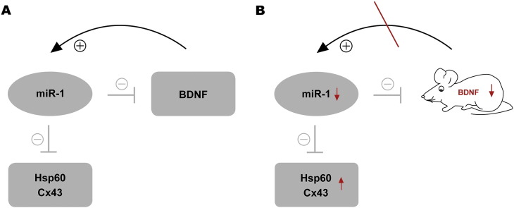
Conclusions: Our results indicate that deletion of Bdnf in DRG neurons leads to a temporary dysregulation of miR-1, suggesting an impairment of a presumable feedback loop between BDNF protein and its targeting miR-1. This appears to affect its downstream protein Hsp60 and as a consequence might influence the phenotype after inducible Bdnf gene deletion. While this appears to be a MEF2a-/SRF-independent and transient effect, expression levels of various other miRNAs may remain permanently altered.

Keywords: BDNF-Advillin-Cre-ERT2; Bdnf; Dorsal root ganglion; Gene deletion; Neuropathic pain; miR-1; microRNA.


Figures

Fig. 1
Fig. 1
BDNF expression in adult Bdnf cKO mice at an early time point after BDNF gene deletion. Quantification of BDNF mRNA in (A) DRG, (B) sciatic nerve and (C) spinal cord with qRT-PCR, presented as relative expression levels (n = 9). ELISA measurements of BDNF in (D) DRG, (E) sciatic nerve and (F) spinal cord (n = 5). White bars represent data from WT mice, while black bars represent data from BDNFAvCreERT2 mice 10 days after the last tamoxifen injection. Data are presented as mean ± SEM. Statistical analysis was performed with unpaired t-test; ***p < 0.001; **p < 0.01.
Fig. 2
Fig. 2
miR-1 is downregulated in DRG of adult Bdnf cKO mice at an early time point after BDNF gene deletion. (A) Expression of miR-1 and (B) miR-124a (control) in DRG were detected using qRT-PCR. Data are presented as normalized levels (n = 9). White bars represent data from WT mice, while black bars represent data from BDNFAvCreERT2 mice 10 days after the last tamoxifen injection. Data are presented as mean ± SEM. Statistical analysis was performed with unpaired t-test; *p < 0.05.
Fig. 3
Fig. 3
Hsp60 and Cx43 expression in DRG of adult Bdnf cKO animals at an early time point after BDNF gene deletion (A) Hsp60 and (B) Cx43 protein expression in DRG were determined by Western blot analysis. Data are presented as normalized levels (n = 8). White bars represent data from WT mice, while black bars represent data from BDNFAvCreERT2 mice 10 days after the last tamoxifen injection. Data are presented as mean ± SEM. Statistical analysis was performed with unpaired t-test; *p < 0.05.
Fig. 4
Fig. 4
BDNF expression in DRG of adult Bdnf cKO mice at a late time point after BDNF gene deletion. Quantification of BDNF mRNA in DRG with qRT-PCR, presented as normalized levels (n = 5). White bars represent data from WT mice, while black bars represent data from BDNFAvCreERT2 mice 32 days after the last tamoxifen injection. Data are presented as mean ± SEM. Statistical analysis was performed with unpaired t-test; *p < 0.05.
Fig. 5
Fig. 5
MiR-1 expression changes in DRG of adult Bdnf cKO mice at a late time point after BDNF gene deletion. Expression of miR-1 in DRG using qRT-PCR. Data are presented as normalized levels (n = 5). White bars represent data from WT mice, while black bars represent data from Bdnf cKO mice 32 days after the last tamoxifen injection. Data are presented as mean ± SEM. Statistical analysis was performed with unpaired t-test.
Fig. 6
Fig. 6
Hsp60 and Cx43 expression in DRG of adult Bdnf cKO animals at a late time point after BDNF gene deletion. Hsp60 (A) and Cx43 (B) protein expression in DRG were determined by Western blot analyses. Data are presented as normalized levels (n = 5). White bars represent data from WT mice, while black bars represent data from Bdnf cKO mice 32 days after the last tamoxifen injection. Data are presented as mean ± SEM. Statistical analysis was performed with unpaired t-test.
Fig. 7
Fig. 7
MiRNA microarray in DRG of adult Bdnf cKO mice at a late time point after BDNF gene deletion. (A) Hierarchical clustering of differentially expressed microRNAs. 167 miRNAs were significantly dysregulated in DRG of late targeted adult Bdnf cKO mice (p < 0.05). Signal intensities (log2, normalized) are color coded: Green denotes downregulation while red denotes upregulation relative to the median. Statistical analysis was performed using one-way-ANOVA. (B) 54 differentially expressed miRNAs in DRG of late targeted adult Bdnf cKO mice. The shown data required a minimum miRNA expression change of 20% compared to WT mice and are grouped by statistical significance (*p < 0.05; **p < 0.01). Statistical analysis was performed with one-way ANOVA.
Fig. 8
Fig. 8
Model of miR-1-associated effects in DRG-specific conditional BDNF-deficient mice. (A) miR-1 negatively regulates BDNF and the neuropathic pain-associated proteins Hsp60 and Cx43. Hypothesized positive regulatory loop from BDNF towards miR-1. (B) Hypothesized impairment of the positive feedback loop due to BDNF gene targeting.

Similar articles

Cited by

References

    1. Bartel D.P. MicroRNAs: genomics, biogenesis, mechanism, and function. Cell. 2004;116:281–297. - PubMed
    1. Bastian I., Tam Tam S., Zhou X.-F.F., Kazenwadel J., Van der Hoek M., Michael M.Z., Gibbins I., Haberberger R.V. Differential expression of microRNA-1 in dorsal root ganglion neurons. Histochem. Cell Biol. 2011;135:37–45. - PubMed
    1. Bolstad B.M., Irizarry R.A., Astrand M., Speed T.P. A comparison of normalization methods for high density oligonucleotide array data based on variance and bias. Bioinformatics. 2003;19:185–193. - PubMed
    1. Brandenburger T., Grievink H., Heinen N., Barthel F., Huhn R., Stachuletz F., Kohns M., Pannen B., Bauer I. Effects of remote ischemic preconditioning and myocardial ischemia on microRNA-1 expression in the rat heart in vivo. Shock. 2014;42:234–238. - PubMed
    1. Chen J.-F.F., Mandel E.M., Thomson J.M., Wu Q., Callis T.E., Hammond S.M., Conlon F.L., Wang D.-Z.Z. The role of microRNA-1 and microRNA-133 in skeletal muscle proliferation and differentiation. Nat. Genet. 2006;38:228–233. - PMC - PubMed