. 2016 Jul 12;16(2):531-544.
doi: 10.1016/j.celrep.2016.05.093. Epub 2016 Jun 23.

Distinct Subunit Domains Govern Synaptic Stability and Specificity of the Kainate Receptor

Affiliations

Distinct Subunit Domains Govern Synaptic Stability and Specificity of the Kainate Receptor

Christoph Straub et al. Cell Rep. .

Abstract

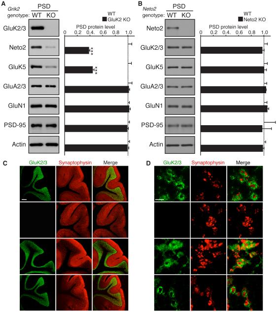
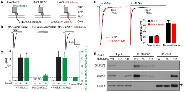
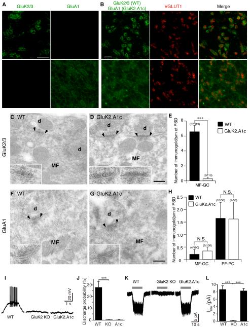
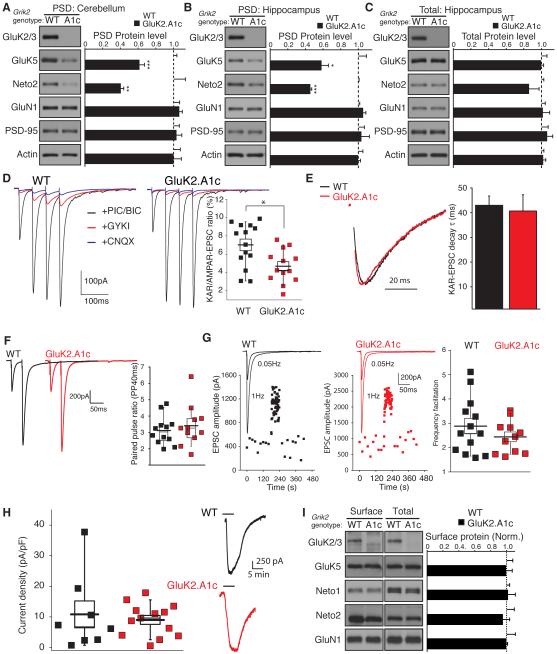
Synaptic communication between neurons requires the precise localization of neurotransmitter receptors to the correct synapse type. Kainate-type glutamate receptors restrict synaptic localization that is determined by the afferent presynaptic connection. The mechanisms that govern this input-specific synaptic localization remain unclear. Here, we examine how subunit composition and specific subunit domains contribute to synaptic localization of kainate receptors. The cytoplasmic domain of the GluK2 low-affinity subunit stabilizes kainate receptors at synapses. In contrast, the extracellular domain of the GluK4/5 high-affinity subunit synergistically controls the synaptic specificity of kainate receptors through interaction with C1q-like proteins. Thus, the input-specific synaptic localization of the native kainate receptor complex involves two mechanisms that underlie specificity and stabilization of the receptor at synapses.


Figures

Figure 1
Figure 1. Synaptic localization of KARs is determined by GluK2, but not GluK5 or Neto2, in cerebellar granule cells
Distribution of components of KAR complex in the cerebellum of the indicated knockout (KO) mice. (A, B) Protein levels of GluK5 and Neto2 were reduced in the cerebellar PSD-enriched fraction of GluK2 KO mice (A), but unaltered in Neto2 KO mice (B). Protein levels of AMPAR (GluA2/3), NMDAR (GluN1), PSD-95, and actin were unaltered (n = 4 each). (C) GluK2/3 signal was not detected in the granular layer of GluK2 KO mice, but was detected in Neto2 and GluK5 KO mice. (D) High-magnification images of cerebellar glomeruli. No obvious change in GluK2 distribution was observed in Neto2 and GluK5 KO mice. Synaptophysin is a presynaptic marker. Scale bars: 200 µm (C), 5 µm (D). GL, PCL, and ML designate granular, Purkinje cell, and molecular layers, respectively. Data in A and B are given as mean ± s.e.m.; ***, P < 0.001 (Student’s t-test).
Figure 2
Figure 2. Roles of the GluK2 cytoplasmic domain in formation of the KAR complex
(A) Schematic diagram of each GluK2 mutant tested. GluK2ΔC indicates deletion of the C-terminal cytoplasmic domain, and GluK2.A1cyto indicates replacement of the cytoplasmic domain of GluK2 with that of the GluA1 AMPAR subunit. Epitopes for anti GluK2/3 or GluA1-antibodies (Ab) are indicated. NTD: N-terminal domain; LBD: ligand-binding domain; TMD: trans-membrane domain; cyto: cytoplasmic C-terminus. (B, C) Glutamate-evoked currents and surface expression were measured by two-electrode voltage-clamp recording and chemiluminescence assay in oocytes injected with various cRNAs, as indicated. (B) Representative traces are shown; gray bar indicates bath application of glutamate (1 mM). (C) Quantitation of peak amplitudes of glutamate-evoked currents (black) and surface expression of HA-tagged GluK2 (green) (n = 10 each). Deletion of the GluK2 cytoplasmic domain abolished surface expression, and replacing the cytoplasmic domain of GluK2 with that of GluA1 restored both surface expression and activity. Green dashed line indicates the background level, defined as the signal from un-injected oocytes. N.D.: not detectable. (D) Responses to 2 ms or 300 ms applications (bars) of 1 mM glutamate in outside-out oocyte membrane patches expressing GluK2 (black) or GluK2.A1cyto (red). Bar graph showing the mean weighted time constants of deactivation and desensitization from bi-exponential fits to the decay of currents. (E) GluK5 interaction with GluK2 or GluK2.A1cyto was analyzed by co-immunoprecipitation using cerebral cortical lysate from wild-type (WT), GluK2 knockout (KO), and GluK2.A1c KI mice (A1c), using antibodies shown in (A). GluK2.A1cyto was detected weakly at a slightly higher molecular weight than that of endogenous GluA1 (arrow). Total GluK5 expression was reduced in GluK2 KO, but not in GluK2A1c KI mice (Input). Data are shown as mean ± s.e.m.
Figure 3
Figure 3. The GluK2 cytoplasmic domain is required for synaptic KAR localization in cerebellar granule cells
(A-H) Distribution of KARs in cerebellum of GluK2.A1c KI mice. (A) Immunohistochemical staining of the granular cell layer of mouse cerebellum. GluK2/3 signal was observed only wild-type (WT) mice, whereas GluA1 signal was observed only in GluK2.A1cyto KI mice. Because of no endogenous GluA1 in the granule cells, the GluA1 signal indicates specific expression of GluK2.A1cyto protein. (B) GluK2 was enriched at cerebellar glomeruli around the mossy fiber presynaptic marker VGLUT1, whereas GluK2.A1cyto was distributed diffusely. Scale bars: 10 µm. (C-H) Immuno-electron microscopic images of GluK2 and GluK2.A1cyto proteins. Inserts show high-magnification of labeled synapses. Scale bars: 200 nm. (C–E) GluK2 was detected at MF-GC synapses in WT mice, but not in GluK2.A1c KI mice. (F–H) No GluK2.A1cyto signal was detected at MF-GC synapses from both WT and GluK2.A1c KI mice with anti-GluA1 C-terminal antibody. By contrast, endogenous GluA1 was detected at similar levels in cerebellar parallel fiber (PF)-Purkinje cell (PC) synapses in both WT and GluK2.A1c KI mice. Numbers of immunogold-labeled synapses and total analyzed synapses are indicated in parentheses. (I-L) KAR activity was measured in cerebellar granule cells. To isolate KAR activity from other glutamate receptors, recordings were performed on the stargazer genetic background. (I, J) Mossy fiber-evoked responses were recorded under the whole-cell current-clamp configuration. KAR-dependent synaptic transmission at cerebellar mossy fiber–granule cell synapses was abolished in both GluK2 KO and GluK2A1c KI mice (WT n =5, KO and K2.A1c n = 4 each). (K, L) KAR activity at the cell surface was measured using 300 µM glutamate (gray bar) in the presence of 100 µM picrotoxin and 100 µM D-AP5. Surface KAR activity was detected at similar levels in WT and GluK2.A1cyto KI mice, but not in GluK2 KO (GluK2−/−) mice (WT n =5, KO and A1c n = 4 each). Data are given as mean ± s.e.m. ***, P < 0.001 (Student’s t-test).
Figure 4
Figure 4. Selective reduction in synaptic KARs in GluK2.A1c KI mice
(A) PSD-enriched fractions were purified from cerebella of wild-type (WT) and GluK2.A1c KI mice. GluK5 levels in the cerebellar PSD-enriched fraction was reduced in GluK2.A1c KI mice. (B and C) Protein levels in the PSD-enriched fraction (B) and total (C) were measured in hippocampi from WT and GluK2.A1c KI mice (n = 6). KAR components GluK5 and Neto2 were specifically reduced in the hippocampal PSD fraction, without changes in the total protein levels. Levels of other excitatory synaptic proteins (GluA1, GluN1 and PSD-95) were unaltered. (D–G) Synaptic activity at hippocampal mossy fiber–CA3 pyramidal cell synapses were measured under the whole-cell voltage-clamp configuration (Vh = −70 mV) in acute slices. (D) EPSCs were measured with combinations of various blockers following four consecutive stimulations of mossy fibers. EPSCs were isolated by addition of picrotoxin and bicuculline. KAR-mediated EPSCs were isolated as currents insensitive to 50 µ M GYKI53655 and sensitive to 10 µ M CNQX. The ratio of KAR-mediated to AMPAR-mediated EPSCs (the difference between total EPSCs and KAR-mediated EPSCs) was significantly reduced in GluK2.A1c KI mice (n = 13) relative to that in WT mice (n = 14). (E) No significant changes in the decay kinetics of KAR-mediated EPSCs were observed (WT n =13; K2.A1c n = 11). (F) Paired-pulse ratio of AMPAR-mediated EPSCs with a 40-ms interval did not differ (WT n =12; K2.A1c n = 11). (G) Frequency facilitation was unchanged (WT n =13; K2.A1c n = 11). (H) Kainate-evoked current density measured in CA3 pyramidal cells and two representative traces from mutant and wild-type mice (I) Surface expression of proteins in acute hippocampal slices was measured using cell-impermeable Sulfo-NHS-SS-biotin. No changes in GluK5, Neto1 and Neto2 as well as GluN1 were observed in the “Surface” and “Total” fractions between WT and GluK2.A1c KI mice (n = 4). Data are given as mean ± s.e.m.; * P < 0.05, ** P < 0.01, *** P < 0.005 (Student’s t-test).
Figure 5
Figure 5. Distinct mechanisms for synaptic stabilization and synapse specificity of KARs in hippocampus
KAR distribution was examined in hippocampus from wild-type (WT) and GluK2.A1c KI; GluA1 KO double-mutant mice. (A, B) GluK2 and GluK2.A1cyto (A) as well as GluK5 (B) were observed at the stratum lucidum in WT and GluK2.A1c KI; GluA1 KO double-mutant mice, respectively. Scale bar: 100 µ m.
Figure 6
Figure 6. High-affinity GluK4/5 subunits mediate synapse specificity of KARs in the hippocampus
The distribution of KAR components in the hippocampus was examined by immunohistochemistry and biochemical fractionation. (A and B) Immunostaining of hippocampal sections without pepsin treatment (see Experimental Procedures) revealed reduction in the GluK2/3 signal at the stratum lucidum (SL) in GluK4/5 double-knockout (DKO) mice. On the other hand, GluK2/3 signal at the stratum radiatum (SM) was elevated in GluK4/5 DKO mice. (C) GluK2/3 distribution in the dentate gyrus was unaltered (G; ML = molecular layer, GCL = granular layer). (D, E) Immunostaining of hippocampal sections after pepsin treatment. In wild-type mice (WT), a strong GluK2/3 signal was detected at the stratum lucidum, but not at the stratum radiatum or stratum pyramidale (SP). This highly compartmentalized pattern was abolished in GluK4/5 DKO mice, but was preserved in Neto1/2 DKO with a slight increase in the GluK2/3 signal at the SP. Images represent GluK2/3 localization at lower (D) and higher (E) magnifications. Scale bars: 100 μm (A, D), 50 μm (E), 25 μm (B, C). (F, G) Protein levels in the PSD fraction (F) and total (G) were measured in hippocampus (n = 5). Protein levels of KAR components (GluK2/3 and Neto1) were significantly reduced in the PSD fraction of GluK4/5 DKO, but total expression was unaltered. (H, I) Protein levels in the PSD fraction (H) and total (I) were measured in hippocampus (n = 3-4). Protein levels of KAR component (Neto1) were further reduced in the PSD fraction of GluK2.A1c KI; GluK4/5 DKO triple-mutant mice, but total expression was unaltered. (J, K) Immuno-electron microscopic images of Neto1 protein. PSDs are indicated by arrowheads. Inserts show high-magnification of labeled synapses. Scale bars = 200 nm (J) Neto1 was detected at hippocampal MF-CA3 synapses in WT mice, but not in Neto1 KO mice. On the other hand, Neto1 was reduced in GluK2.A1c KI mice. No Neto1 signal was detected in GluK4/5 DKO and GluK2.A1c KI; GluK4/5 DKO triple-mutant mice. (K) GluK2.A1cyto signal detected by anti GluA1 antibody was detected in GluK2.A1c KI; GluA1 KO double-mutant mice, but not in GluK2.A1c KI; GluK4/5 DKO, GluA1 KO quadruple-mutant mice. Numbers of immunogold-labeled synapses and total analyzed synapses are indicated in parentheses. Data are given as mean ± s.e.m. *, P < 0.05; ** P < 0.01; ***, P < 0.001 (Student’s t-test).
Figure 7
Figure 7. The GluK5 extracellular domain mediates synapse specificity
(A) Immunostaining of hippocampal sections revealed substantial reduction of GluK2/3 signal in the stratum lucidum layer in GluK4/5 DKO compared to GluK4 KO mice. (B) Schematic diagram of chimeras of GluK5 and GluK2 with GFP at their C-terminus. (C) Surface expression of the extracellularly HA-tagged GluK5-GFP chimeras in cRNA-injected oocytes was measured using chemiluminescence assay. HA-K5-GFP alone did not express at the cell surface. On the other hand, GluK2 co-expression enhanced surface expression of HA-GluK5-GFP, HA-GluK5.K2extra-GFP and HA-GluK5.K2cyto-GFP, but not HA-GluK5.K2TM-GFP (n = 6-8). Expression of chimeric proteins was confirmed by western blotting. (D) Upon stereotaxic injection of AAV carrying GluK5-GFP, GluK5-GFP signal was observed in AAV-injected hemispheres in GluK4/5 DKO hippocampus (top). Re-introducing GluK5-GFP and GluK5.K2cyto-GFP into GluK4/5 DKO restored the stratum lucidum localization of endogenous GluK2 (Magenta), whereas GluK5.K2extra-GFP failed. Composite images were shown. (E) HA-tagged C1QL3/nCLP3 bound to the GluK5 extracellular domain tagged with human Fc domain (GluK5extra-Fc). Two proteins expressed independently were mixed and pulled down with protein A-sepharose. HA-C1QL3 was pulled down with GluK5extra-Fc strongly, but not with bovine serum albumin (control). Addition of calcium (Ca2+) was required for their interaction. (F) Immunostaining of C1QL2/nCLP2 in the hippocampus resulted in a selective distribution at the stratum lucidum, mimicking the distribution pattern of KARs. (G) The stratum lucidum distribution of C1QL2 was markedly reduced in GluK4/5 DKO mice. Data in C are given as mean ± s.e.m. ** P < 0.01; ***, P < 0.001. Scale bars: 100 μm (A, left panels; F, G), 20 μm (A, right panel; D, bottom).

References

    1. Bats C, Groc L, Choquet D. The interaction between Stargazin and PSD-95 regulates AMPA receptor surface trafficking. Neuron. 2007;53:719–734. - PubMed
    1. Bolliger MF, Martinelli DC, Sudhof TC. The cell-adhesion G protein-coupled receptor BAI3 is a high-affinity receptor for C1q-like proteins. Proc Natl Acad Sci U S A. 2011;108:2534–2539. - PMC - PubMed
    1. Carta M, Opazo P, Veran J, Athane A, Choquet D, Coussen F, Mulle C. CaMKII-dependent phosphorylation of GluK5 mediates plasticity of kainate receptors. EMBO J. 2013;32:496–510. - PMC - PubMed
    1. Castillo PE, Malenka RC, Nicoll RA. Kainate receptors mediate a slow postsynaptic current in hippocampal CA3 neurons. Nature. 1997;388:182–186. - PubMed
    1. Chen L, Chetkovich DM, Petralia RS, Sweeney NT, Kawasaki Y, Wenthold RJ, Bredt DS, Nicoll RA. Stargazin regulates synaptic targeting of AMPA receptors by two distinct mechanisms. Nature. 2000;408:936–943. - PubMed

LinkOut - more resources