. 2016 Jun 27:6:28112.
doi: 10.1038/srep28112.

Histone modifications and p53 binding poise the p21 promoter for activation in human embryonic stem cells

Affiliations

Histone modifications and p53 binding poise the p21 promoter for activation in human embryonic stem cells

Yoko Itahana et al. Sci Rep. .

Abstract

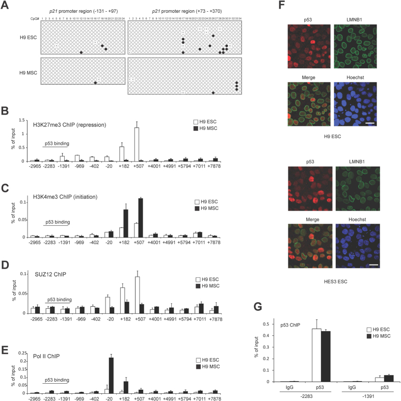
The high proliferation rate of embryonic stem cells (ESCs) is thought to arise partly from very low expression of p21. However, how p21 is suppressed in ESCs has been unclear. We found that p53 binds to the p21 promoter in human ESCs (hESCs) as efficiently as in differentiated human mesenchymal stem cells, however it does not promote p21 transcription in hESCs. We observed an enrichment for both the repressive histone H3K27me3 and activating histone H3K4me3 chromatin marks at the p21 locus in hESCs, suggesting it is a suppressed, bivalent domain which overrides activation by p53. Reducing H3K27me3 methylation in hESCs rescued p21 expression, and ectopic expression of p21 in hESCs triggered their differentiation. Further, we uncovered a subset of bivalent promoters bound by p53 in hESCs that are similarly induced upon differentiation in a p53-dependent manner, whereas p53 promotes the transcription of other target genes which do not show an enrichment of H3K27me3 in ESCs. Our studies reveal a unique epigenetic strategy used by ESCs to poise undesired p53 target genes, thus balancing the maintenance of pluripotency in the undifferentiated state with a robust response to differentiation signals, while utilizing p53 activity to maintain genomic stability and homeostasis in ESCs.


Figures

Figure 1
Figure 1. p21 expression is suppressed in human embryonic stem cells.
(A) p21 expression is suppressed in hESCs and hiPSCs compared to hMSCs. Protein lysates from the indicated cells were analyzed by Western blotting with the indicated antibodies. The passage number is shown in brackets. 70 μg of protein lysate was loaded in each lane. (B) p21 expression in hESCs is about 50 times lower than in hMSCs, as analyzed by Western blotting with the indicated antibody. 150 μg of protein lysate from H9 hESCs was loaded in lane 1. The amount of total protein lysate loaded relative to hESC is indicated. (C) p21 mRNA levels are lower in H9 hESCs than in hMSCs, as assessed by qRT-PCR (n = 3, means ± SD). The mean value of mRNA expression in H9 hESCs is set at 1, and relative expression is shown. TBP was used as an internal control for normalization. (D,E) p53 is required for p21 expression in H9 hMSCs (passage number 8). H9 hMSCs were transfected with control and p53 siRNAs. p21 protein levels (D) were analyzed by Western blotting. 50 μg of protein lysate was loaded in each lane. p21 mRNA levels (E) were analyzed as in (C). The mean value of mRNA expression in control siRNA transfected cells is set at 1, and relative expression is shown. (F,G) p21 expression in H9 hESCs remains very low upon p53 activation by DNA damage. H9 hESCs and H9 hMSCs were treated with the indicated concentration of etoposide (F) or hydroxyurea (G) for 24 hrs and harvested for Western blotting. The passage numbers of H9 hESCs and hMSCs are P37 and P10 respectively. 50 μg of protein lysate was loaded in each lane.
Figure 2
Figure 2. Protein stability and translational efficiency do not underlie low levels of p21 protein in hESCs.
(A) The half-life of the p21 protein is comparable in H9 hESCs and hMSCs. Cells were treated with cycloheximide and cell lysates were harvested at the indicated time for Western blotting analysis. An increased amount (250 μg) of lysate and longer exposure time were used to enable detection of p21 in hESCs. 50 μg of protein lysate was used for hMSCs. The passage numbers of H9 hESCs and hMSCs are P46 and P5 respectively. (B) The p21 protein is not subject to increased proteasomal degradation in hESCs relative to hMSCs. Cells were treated with MG132 or hydroxyurea, and cell lysates were analyzed by Western blotting. The passage number of H9 hESCs and hMSCs are P46 and P6 respectively. 50 μg of protein lysate was loaded in each lane. (C) Translation state analysis of p21 mRNA in H9 hESCs. Representative polysome profile from H9 hESCs indicating monosomes and polysome fractions is shown in the left panel. qRT-PCR analysis shows the percentage of TBP and p21 mRNA in each fraction in the right panels.
Figure 3
Figure 3. p21 expression in hESCs is repressed by histone H3K27me3.
(A) p21 promoter DNA is not significantly methylated in H9 hESCs and hMSCs. The CpG rich promoter regions of p21 (−131− + 97 bp and +73 − + 370 bp in relation to the transcriptional start site, TSS) were subjected to bisulfite sequencing. Each line shows independent sequencing results (n = 8–11). Filled circles indicate methylated CpGs and open circles represent unmethylated CpGs (X denotes un-identified nucleotide sequence). Clones with at least 90% cytosine conversion were considered as unmethylated sites. (B–E) ChIP analysis on the p21 gene locus was performed using protein lysates of H9 hESC and hMSCs with the indicated antibodies. ChIP-enriched DNA was quantified by qPCR (n = 3, mean ± SD). Values are shown as a percentage of input DNA. The amplicon’s name indicates the position of the central base pair of the amplicon relative to the TSS of p21 gene. Note that p21 gene locus is silenced by H3K27me3. (F) p53 localizes in the nucleus. Immunofluorescence staining of p53 and LMNB1 (nuclear envelope marker) in hESC line H9 and HES3. Scale bar, 20 μm. (G) p53 protein binds equally well to the p21 promoter in H9 hESC and hMSC. ChIP analysis with p53 antibody was performed as in (B). Mouse IgG was used as the negative control for immunoprecipitation.
Figure 4
Figure 4. DZNep rescues p21 expression in hESCs, and p21 expression in hESCs induces differentiation.
(A,B) DZNep reduces H3K27me3 marks on the p21 locus without affecting p53 binding to the p21 promoter. H9 hESCs were treated with DZNep (1 μM) for 2 days and ChIP analysis was performed using p53 antibody (A) or H3K27me3 antibody (B) as in Fig. 3 (n = 3, mean ± SD). **p < 0.01, two-tailed unpaired t-test compared to control cells. The passage number of H9 hESCs is P35. (C) DZNep induces the expression of p21 in hESCs. H9 hESCs and hMECs were treated with DZNep at the indicated dose for 2 days. Proteins were harvested for Western blotting analysis with the indicated antibodies. The passage numbers of H9 hESCs and hMSCs are P35 and P9 respectively. (D) Ectopic p21 expression in hESCs. H9 hESCs were infected with retrovirus carrying either empty (pMSCV) or p21 and selected with blasticidin for 3 days. Immunofluorescence staining of p21 and differentiation marker, LMNA are shown. Scale bar, 20 μm. (E) Introduction of p21 reduces the expression of pluripotency markers and increases the differentiation markers in H9 hESCs. The mRNA expression of the indicated genes was analyzed as in Fig. 1C. The mean value of mRNA expression in pMSCV infected cells is set at 1, and relative expression is shown. **p < 0.01, ***p < 0.001, two-tailed unpaired t-test compared to control cells. (F) Expression of p21 in hESCs triggers a differentiated cell morphology (Phase) and reduces alkaline phosphatase staining (AP staining). Scale bar, 100 μm.
Figure 5
Figure 5. A subset of p53 target genes is subject to epigenetic silencing in hESCs.
(A) H3K4me3 and H3K27me3 levels at the TSSs of putative p53 target genes in H1 hESCs were plotted (left). The same set of genes were also plotted for H3K4me3 and H3K27me3 levels in H1-derived hMSCs (right). The p21 gene is marked in black open circle and shows high levels of H3K27me3 in hESCs, indicating that it is epigenetically repressed. The color corresponds to the mRNA expression level (FPKM) for each gene in the respective cell type. (B) Heatmap showing relative mRNA expression of p53 target gene candidates in H1 hESCs and hMSCs. Genes were sorted by the ratio of H3K27me3 levels in hESCs:hMSCs. FPKM values were normalized per gene to obtain relative expression values. The p21 gene and the genes selected for further analysis in Fig. 5C are indicated in red. (C) A subset of p53 target gene candidates is suppressed in H9 hESCs and requires p53 for expression in hMSCs. RNA was extracted from H9 hESCs and hMSCs and the indicated genes were analyzed by qRT-PCR (n = 3, means ± SD) (left plot). The mean value of mRNA expression in H9 hESCs is set at 1, and relative expression is shown in the left plot. H9 hMSCs were transfected with control and p53 siRNAs, and mRNA levels of each gene were analyzed similarly (right plot). The mean value of mRNA expression in control siRNA transfected H9 MSCs is set at 1, and relative expression is shown in the right plot. The passage number of H9 hMSCs is P8. (D) A subset of known p53 target genes is expressed in a p53-dependent manner in mouse ESCs. RNA was extracted from mouse p53+/− ESCs and littermate p53−/− ESCs, and the indicated genes was analyzed as in (C). The mean value of mRNA expression in p53+/− ESCs is set at 1, and relative expression is shown. (E) A conclusion model: A subset of p53 target genes such as p21 is poised but silenced by bivalent promoters in hESCs, and activated by releasing H3K27me3 marks upon differentiation.

References

    1. Song H., Chung S. K. & Xu Y. Modeling disease in human ESCs using an efficient BAC-based homologous recombination system. Cell Stem Cell 6, 80–89 (2010). - PubMed
    1. Lee K. H. et al.. A genomewide study identifies the Wnt signaling pathway as a major target of p53 in murine embryonic stem cells. Proc. Natl. Acad. Sci. USA 107, 69–74 (2010). - PMC - PubMed
    1. Han M. K. et al.. SIRT1 regulates apoptosis and Nanog expression in mouse embryonic stem cells by controlling p53 subcellular localization. Cell Stem Cell 2, 241–251 (2008). - PMC - PubMed
    1. Lin T. et al.. p53 induces differentiation of mouse embryonic stem cells by suppressing Nanog expression. Nat. Cell Biol. 7, 165–171 (2005). - PubMed
    1. Maimets T., Neganova I., Armstrong L. & Lako M. Activation of p53 by nutlin leads to rapid differentiation of human embryonic stem cells. Oncogene 27, 5277–5287 (2008). - PubMed

Publication types

MeSH terms

Substances