. 2016 Jun 24:16:50.
doi: 10.1186/s12935-016-0327-0. eCollection 2016.

Involvement of human chorionic gonadotropin in regulating vasculogenic mimicry and hypoxia-inducible factor-1α expression in ovarian cancer cells

Affiliations

Involvement of human chorionic gonadotropin in regulating vasculogenic mimicry and hypoxia-inducible factor-1α expression in ovarian cancer cells

Min Su et al. Cancer Cell Int. .

Abstract

Background: Human chorionic gonadotropin (hCG) can play a crucial role in angiogenesis. In the present study, we focused on hCG to gain insight into its potential effects on vasculogenic mimicry (VM) in ovarian cancer cells.

Methods: Ovarian cancer OVCAR-3 cells were incubated with different concentrations of recombinant hCG in 3-dimensional cultures. VM was identified by morphological observations and vascular endothelial cell marker detection in OVCAR-3 cells. Expression of hCG, hypoxia-inducible factor-1α (HIF-1α), and the endothelial cell markers CD31, VEGF, and factor VIII were detected by reverse transcription polymerase chain reaction and western blotting. The effect of hCG on endothelial cell-marker expression in ovarian cancer cells was further explored using small interfering RNA (siRNA) and plasmid-based approaches.

Results: Incubation of OVCAR-3 cells with recombinant hCG induced vessel-like network formation, which was accompanied by significant elevation of vascular marker expression. Attenuation of hCG expression by siRNA in OVCAR-3 cells suppressed the expression of endothelial cell markers and HIF-1α by tumour cells. Overexpression of hCG in OVCAR-3 cells resulted in increased expression of endothelial cell markers and HIF-1α.

Conclusions: HCG was crucial for changing the phenotype of OVCAR-3 cells to endothelial-like cells. The effect of hCG induction on VM in ovarian cancer cells is potentially associated with HIF-1α.

Keywords: Human chorionic gonadotropin; Hypoxia inducible factor-1α; Ovarian cancer; Vasculogenic mimicry.


Figures
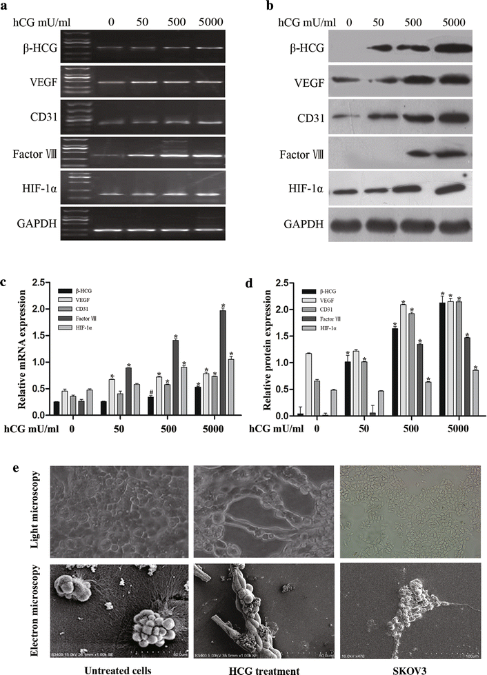
Fig. 1
Fig. 1
Expression of vascular cell markers and hCG in and morphological flexibility of hCG-treated OVCAR-3 cells. a, b Expression levels of vascular markers CD31, VEGF, factor VIII, hCG, and HIF-1α were determined in OVCAR-3 cells exposed to 50, 500, or 5000 mU/ml hCG for 7 days. HCG treatment stimulated the expression of vascular markers and HIF-1α in OVCAR-3 cells in a dose-dependent manner. a The mRNA levels were analysed by RT-PCR. b Protein levels were detected by western blotting. c, d Band densities were quantified by densitometric analysis. Protein and mRNA content was quantified for 3 independent replicates and the data are presented as the mean ± SD. The data shown are presented after normalization with GAPDH expression and were analysed using 1-way ANOVA. *p < 0.01, #p < 0.05. e Light and scanning-electron microscopy observations showed tubular network and channel formation by OVCAR-3 cells in the 3D matrix after exposure to 5000 mU/ml hCG. Representative morphological changes are shown
Fig. 2
Fig. 2
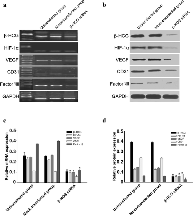
Inhibition of hCG expression using siRNA resulted in suppressed vascular marker and HIF-1α expression. a, b Expression of hCG in OVCAR-3 cells was inhibited by siRNA targeting hCG mRNA, but not by a negative control siRNA. Compared with untransfected OVCAR-3 cells and mock-transfected OVCAR-3 cells, the expression of CD31, VEGF, factor VIII, and HIF-1α decreased in OVCAR-3 cells transfected with hCG siRNA. a mRNA expression of the vascular cell marker, HIF-1α and hCG was analysed by RT-PCR. b Protein expression of the vascular cell marker, HIF-1α and hCG was analysed by western blotting. c, d Band densities were quantified by densitometric analysis. Protein and mRNA content measured in 3 independent replicates was quantified and the data are presented as the mean ± SD. The data shown are presented after normalization with GAPDH bands and analysed by 1-way ANOVA. *p < 0.01
Fig. 3
Fig. 3
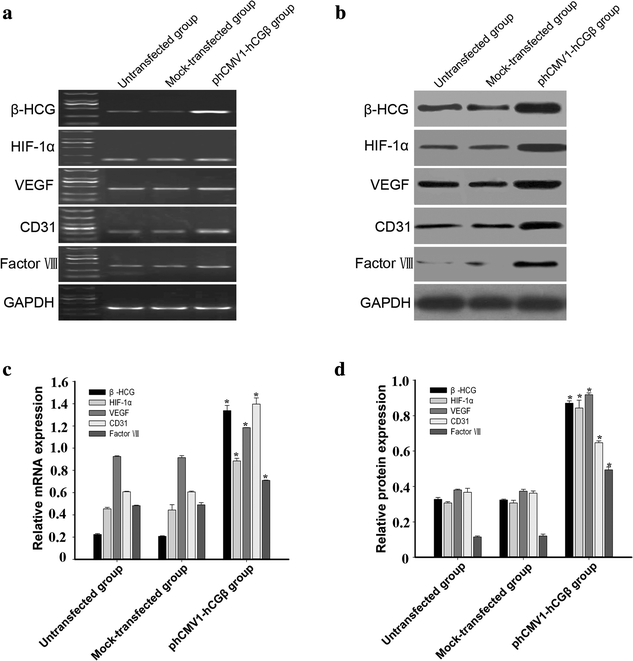
Up-regulated expression of vascular markers and HIF-1α in OVCAR-3 cells transfected with the phCMV1 vector expressing β-hCG (phCMV1-hCGβ). a, b HCG expression increased significantly in transfected OVCAR-3 cells, as determined by RT-PCR and western blot analysis. Compared with untransfected and mock-transfected cells, both mRNA (a) and protein expression (b) of CD31, VEGF, Factor VIII, and HIF-1α increased significantly. c, d Band densities were quantified by densitometric analysis. Protein and mRNA content measured in 3 independent replicates was quantified and the data are presented as the mean ± SD. The data shown were normalized to GAPDH bands and analysed by 1-way ANOVA. *p < 0.01
Fig. 4
Fig. 4
Expression of the hCG receptor (hCG-R) in the ovarian cancer cell line OVACR-3. a Confocal image of OVCAR-3 cells following immunofluorescence staining with an hCG-R antibody. Green fluorescence was localized to the periphery of OVCAR-3 cells. Blue fluorescence (PI) was used to demonstrate the nucleus. b, d Different concentrations of hCG did not significantly affect hCG-R expression in OVCAR-3 cells. b Protein expression of hCG-R in OVCAR-3 cells treated with hCG (0, 50, 500, or 5000 mU/ml) for 7 days was detected by western blot analysis. d Expression of hCG-R mRNA in OVCAR-3 treated with hCG (50, 500, or 5000 mU/ml) was detected by RT-PCR. c, e Band densities were quantified by densitometric analysis. The protein and mRNA content measured in 3 independent replicates was quantified and the data are presented as the mean ± SD. The data shown were normalized to GAPDH bands and analysed using 1-way ANOVA. p > 0.05

Similar articles

Cited by

References

    1. Maniotis AJ, Folberg R, Hess A, Seftor EA, Gardner LM, Pe’er J, et al. Vascular channel formation by human melanoma cells in vivo and in vitro: vasculogenic mimicry. Am J Pathol. 1999;155:739–752. doi: 10.1016/S0002-9440(10)65173-5. - DOI - PMC - PubMed
    1. Yao LQ, Feng YJ, Ding JX, Jing HM, Xu CJ, Chen SF, et al. Differentiation of vascular endothelial cells-like derived from epithelial ovarian cancer cells induced by hypoxia. Int J Oncol. 2007;30:1069–1075. - PubMed
    1. Vartanian AA, Stepanova EV, Gutorov SL, Solomko ES, Grigorieva IN, Sokolova IN, et al. Prognostic significance of periodic acid-Schiff-positive patterns in clear cell renal cell carcinoma. Can J Urol. 2009;16:4726–4732. - PubMed
    1. Su M, Yao LQ, Feng YJ, Cheng MJ, Xu CJ, Huang Y, et al. Plasticity of ovarian cancer cell and vasculogenic mimicry in vivo. Int J Gynecol Cancer. 2008;18:476–486. doi: 10.1111/j.1525-1438.2007.01034.x. - DOI - PubMed
    1. Clemente M, Perez M, Illera J, Pena L. Histological, immunohistological, and ultrastructural description of vasculogenic mimicry in canine mammary cancer. Vet Pathol. 2010;47:265–274. doi: 10.1177/0300985809353167. - DOI - PubMed

LinkOut - more resources