

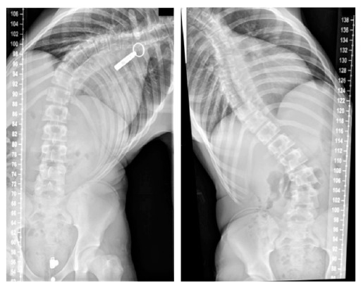
Adolescent Idiopathic Scoliosis
- PMID: 27347243
- PMCID: PMC4897334
- DOI: 10.2174/1874325001610010143
Adolescent Idiopathic Scoliosis
Abstract
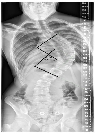
Background: Scoliosis refers to deviation of spine greater than 10 degrees in the coronal plane. Idiopathic Scoliosis is the most common spinal deformity that develops in otherwise healthy children. The sub types of scoliosis are based on the age of the child at presentation. Adolescent idiopathic scoliosis (AIS) by definition occurs in children over the age of 10 years until skeletal maturity.
Objective: The objective of this review is to outline the features of AIS to allow the physician to recognise this condition and commence early treatment, thereby optimizing patient outcome.
Method: A thorough literature search was performed using available databases, including Pubmed and Embase, to cover important research published covering AIS.
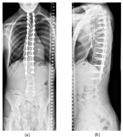
Conclusion: AIS results in higher incidence of back pain and discontent with body image. Curves greater than 50 degrees in thoracic region and greater than 30 degrees in lumbar region progress at a rate of 0.5 to 1 degree per year into adulthood. Curves greater than 60 degrees can lead to pulmonary functional deficit. Therefore once the disease is recognized, effective treatment should be instituted to address the deformity and prevention of its long-term sequelae.
Keywords: Adolescent; deformity; scoliosis; spine.
Figures

References
-
- Le Vay D. The history of orthopaedics: An account of the study and practice of orthopaedics from the earliest times to the modern era. US: Parthenon Publishing; 1990.
-
- Boos N., Aebi M. Spinal disorders: fundamentals of diagnosis and treatment. Germany: Springer; 2008. - DOI
-
- Miller M.D., Thompson S.R., Hart J. Review of orthopaedics. US: Elsevier Health Sciences; 2012.
LinkOut - more resources
Full Text Sources
Other Literature Sources
Medical