. 2016 Sep;11(9):776-82.
doi: 10.1038/nnano.2016.96. Epub 2016 Jun 27.

Three-dimensional mapping and regulation of action potential propagation in nanoelectronics-innervated tissues

Affiliations

Three-dimensional mapping and regulation of action potential propagation in nanoelectronics-innervated tissues

Xiaochuan Dai et al. Nat Nanotechnol. 2016 Sep.

Abstract

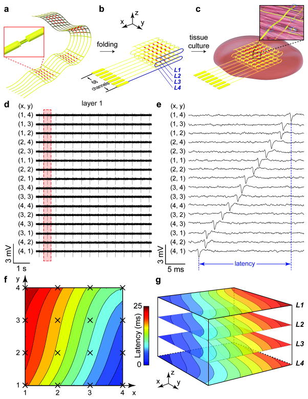
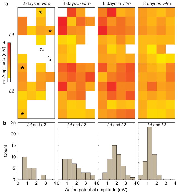
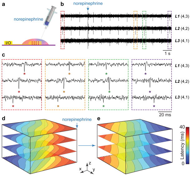
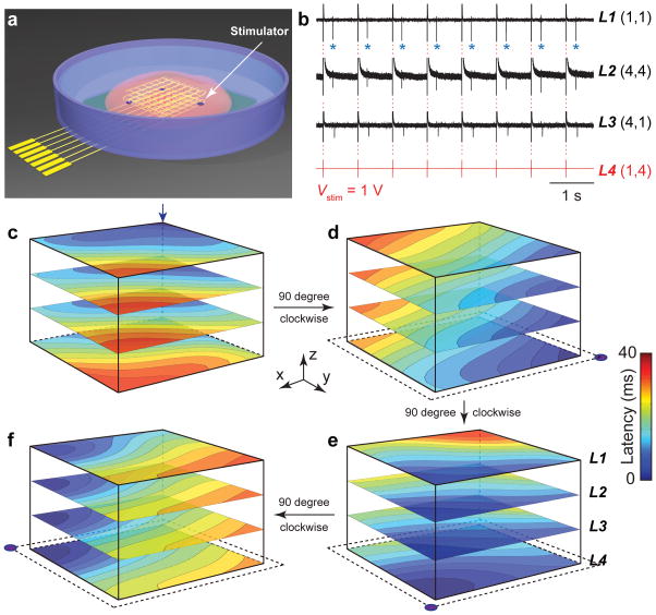
Real-time mapping and manipulation of electrophysiology in three-dimensional (3D) tissues could have important impacts on fundamental scientific and clinical studies, yet realization is hampered by a lack of effective methods. Here we introduce tissue-scaffold-mimicking 3D nanoelectronic arrays consisting of 64 addressable devices with subcellular dimensions and a submillisecond temporal resolution. Real-time extracellular action potential (AP) recordings reveal quantitative maps of AP propagation in 3D cardiac tissues, enable in situ tracing of the evolving topology of 3D conducting pathways in developing cardiac tissues and probe the dynamics of AP conduction characteristics in a transient arrhythmia disease model and subsequent tissue self-adaptation. We further demonstrate simultaneous multisite stimulation and mapping to actively manipulate the frequency and direction of AP propagation. These results establish new methodologies for 3D spatiotemporal tissue recording and control, and demonstrate the potential to impact regenerative medicine, pharmacology and electronic therapeutics.


Conflict of interest statement

The authors declare no competing financial interests.

Figures

Figure 1
Figure 1. 3D spatiotemporal mapping of APs
(a) Schematic of free-standing macroporous nanoelectronic scaffold with nanowire FET arrays (red dots); inset, one nanowire FET. In (ac), a limited number of input/output leads are shown for clarity; the total for the design as indicated in (b) is 68. (b) Folded 3D free-standing scaffolds with four layers of individually addressable FET sensors. (c) Schematic of nanoelectronic scaffold/cardiac tissue resulting from culture of cardiac cells within the 3D folded scaffold; inset, nanoelectronic sensors (blue circles) innervate the 3D cell network. (d) Simultaneous traces recorded from 16 sensors in the top layer (L1) from nanoelectronics-cardiac tissue. The (x,y) coordinates of each element from the 4 × 4 array are shown. (e) Zoom-in view of a single AP spike recorded from each device during the time indicated by the dashed-box in (d). The time latency between APs recorded from different devices is evident and specifically indicated for FETs (4,1) to (1,4). (f) Isochronal map of time latency in L1; mapping area is ca. 25 mm2. (g) 3D isochronal map of time latency through the sample, where L1L4 correspond to the four layers of 4 × 4 device arrays innervating the cardiac tissue. Mapping area is ca. 25 mm2 × 200 μm.
Figure 2
Figure 2. AP evolution during tissue development
(a) Amplitudes of spontaneous extracellular APs recorded from 4 × 6 nanowire FET arrays in two layers at 2, 4, 6 and 8 DIV. White squares correspond to coordinates where extracellular APs are absent or below the detection limit (1× standard deviation of noise level). Time-dependent data recorded from four devices (2 × L1 and 2 × L2) indicated with asterisks at 2 DIV are shown in Supplementary Fig. 5. (b) Histogram of extracellular AP amplitudes recorded from the 3D nanoelectronics-cardiac tissue sample at 2, 4, 6 and 8 DIV.
Figure 3
Figure 3. Arrhythmia induced by localized norepinephrine injection
(a) Schematic of measurement setup highlighting the syringe injection of norepinephrine at a localized spot on the 3D nanoelectronics-cardiac tissue. (b) Time-dependent traces from three sensors in L1, L2, L3 with synchronized and periodic APs. Blue arrow indicates the injection time point of ~25 μL norepinephrine at concentration of 100 μM. (c) Zoom-in view of the four dashed-box regions in (b) depicting time latency between APs before and 5–10 s after norepinephrine addition. (de) 3D isochronal time latency maps before (d) and 5 min after (e) local norepinephrine injection; blue arrow in (d) indicates injection position.
Figure 4
Figure 4. Active spatiotemporal regulation of APs
(a) Schematic illustrating positions of individually addressable stimulator electrodes (purple dots) in the nanoelectronic scaffold. (b) Time-dependent traces recorded from nanowire FETs in layers L1, L2, L3 under periodic biphasic stimulation spike train in L4. Stimulation peak width, amplitude and frequency were 1 ms, 1 V and 1.25 Hz, respectively. Blue asterisks in L1 trace highlight APs (downward spikes) versus capacitive coupling peaks (red dashed-lines). (cf) 3D isochronal time latency maps for original pace-maker foci location (c, blue arrow), and sequential 90 degree rotations of the AP propagation direction using the indicated simulator electrodes (lower corners panels d to f).

References

    1. Langer R, Vacanti JP. Tissue Engineering. Science. 1993;260:920–926. - PubMed
    1. Eschenhagen T, Zimmermann WH. Engineering myocardial tissue. Circ Res. 2005;97:1220–1231. - PubMed
    1. Shimizu T, et al. Fabrication of pulsatile cardiac tissue grafts using a novel 3-dimensional cell sheet manipulation technique and temperature-responsive cell culture surfaces. Circ Res. 2002;90:e40. - PubMed
    1. Dvir T, Timko BP, Kohane DS, Langer R. Nanotechnological strategies for engineering complex tissues. Nat Nanotechnol. 2011;6:13–22. - PMC - PubMed
    1. Papadaki M, et al. Tissue engineering of functional cardiac muscle: molecular, structural, and electrophysiological studies. Am J Physiol-Heart C. 2001;280:168–178. - PubMed

Publication types