

Magnetic Resonance Imaging of Asymptomatic Knees in Collegiate Basketball Players: The Effect of One Season of Play
- PMID: 27347867
- PMCID: PMC5083196
- DOI: 10.1097/JSM.0000000000000283
Magnetic Resonance Imaging of Asymptomatic Knees in Collegiate Basketball Players: The Effect of One Season of Play
Abstract
Objective: To determine the prevalence of abnormal structural findings using 3.0-T magnetic resonance imaging (MRI) in the asymptomatic knees of male and female collegiate basketball players before and after a season of high-intensity basketball.
Design: Institutional review board-approved prospective case series.
Participants: Asymptomatic knees of 24 NCAA Division I collegiate basketball players (12 male, 12 female) were imaged using a 3.0-T MRI scanner before and after the end of the competitive season. Three subjects did not undergo scanning after the season.
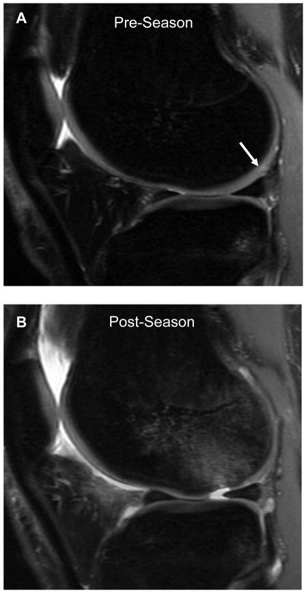
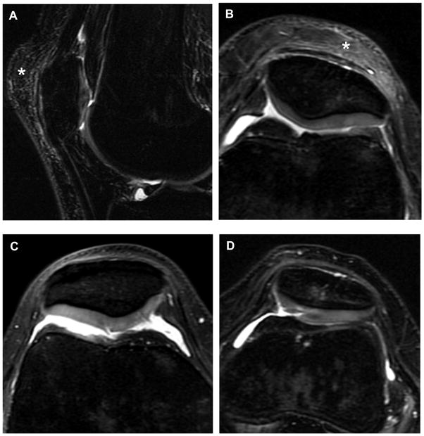
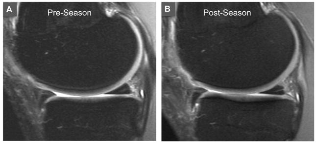
Main outcome measures: Images were evaluated for prepatellar bursitis, fat pad edema, patellar and quadriceps tendinopathy, bone marrow edema, and articular cartilage and meniscal injury.
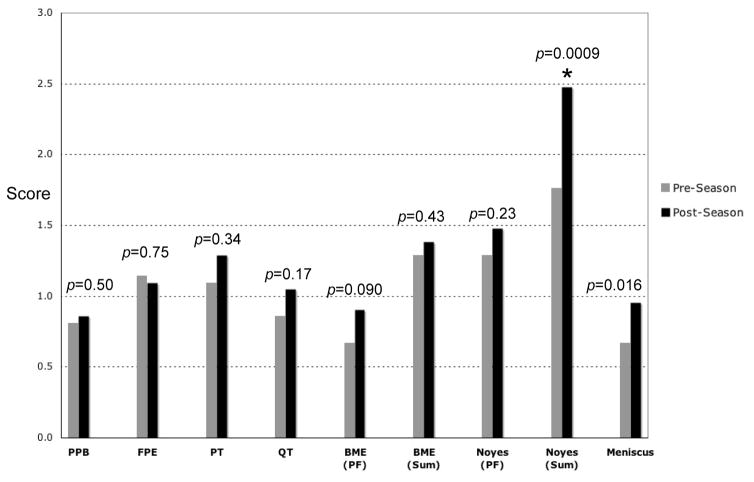
Results: Every knee imaged had at least 1 structural abnormality both preseason and postseason. A high preseason and postseason prevalence of fat pad edema (75% and 81%), patellar tendinopathy (83% and 90%), and quadriceps tendinopathy (75% and 90%) was seen. Intrameniscal signal change was observed in 50% preseason knees and 62% of postseason knees, but no discrete tears were found. Bone marrow edema was seen in 75% and 86% of knees in the preseason and postseason, respectively. Cartilage findings were observed in 71% and 81% of knees in the preseason and postseason, respectively. The cartilage injury score increased significantly in the postseason compared with the preseason (P = 0.0009).
Conclusions: A high prevalence of abnormal knee MRI findings was observed in a population of asymptomatic young elite athletes. These preliminary data suggest that high-intensity basketball may have potentially deleterious effects on articular cartilage.
Figures
Comment in
-
Bedeutung der klinischen Untersuchung im Zusammenhang mit MRT Befunden des Kniegelenks.Sportverletz Sportschaden. 2017 Jan;31(1):12. doi: 10.1055/s-0042-122934. Epub 2017 Mar 30. Sportverletz Sportschaden. 2017. PMID: 28359121 German. No abstract available.
References
-
- Bredella MA, Tirman PF, Peterfy CG, et al. Accuracy of T2-weighted fast spin-echo MR imaging with fat saturation in detecting cartilage defects in the knee: comparison with arthroscopy in 130 patients. Am J Roentgenol. 1999;172(4):1073–80. - PubMed
-
- Potter HG, Linklater JM, Allen AA, et al. Magnetic resonance imaging of articular cartilage in the knee: an evaluation with use of fast spin-echo imaging. J Bone Joint Surg Am. 1998;80(9):1276–84. - PubMed
-
- Kelly MA, Flock TJ, Kimmel JA, et al. MR imaging of the knee: clarification of its role. Arthroscopy. 1991;7(1):78–85. - PubMed
-
- Magee T, Williams D. 3.0-T MRI of meniscal tears. Am J Roentgenol. 2006;187(2):371–5. - PubMed
-
- Ramnath RR, Magee T, Wasudev N, et al. Accuracy of 3-T MRI using fast spin-echo technique to detect meniscal tears of the knee. Am J Roentgenol. 2006;187(1):221–5. - PubMed
MeSH terms
Grants and funding
LinkOut - more resources
Full Text Sources
Other Literature Sources
Medical
