Review
. 2016 Jun 23;8(7):179.
doi: 10.3390/v8070179.

Replication-Competent Influenza A Viruses Expressing Reporter Genes

Affiliations
Review

Replication-Competent Influenza A Viruses Expressing Reporter Genes

Michael Breen et al. Viruses. .

Abstract

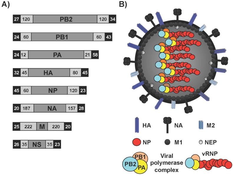
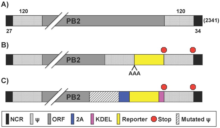
Influenza A viruses (IAV) cause annual seasonal human respiratory disease epidemics. In addition, IAV have been implicated in occasional pandemics with inordinate health and economic consequences. Studying IAV, in vitro or in vivo, requires the use of laborious secondary methodologies to identify virus-infected cells. To circumvent this requirement, replication-competent IAV expressing an easily traceable reporter protein can be used. Here we discuss the development and applications of recombinant replication-competent IAV harboring diverse fluorescent or bioluminescent reporter genes in different locations of the viral genome. These viruses have been employed for in vitro and in vivo studies, such as the screening of neutralizing antibodies or antiviral compounds, the identification of host factors involved in viral replication, cell tropism, the development of vaccines, or the assessment of viral infection dynamics. In summary, reporter-expressing, replicating-competent IAV represent a powerful tool for the study of IAV both in vitro and in vivo.

Keywords: fluorescence; luminescence; plasmid-based reverse genetics; recombinant influenza A virus; replicating-competent reporter-expressing influenza A virus; reporter genes; virus rescue approaches.


Figures

Figure 1
Figure 1
Influenza A virus (IAV) genome organization and virion structure. (A) Genome organization: The eight single-stranded, negative-sense, viral (v)RNA segments PB2, PB1, PA, HA, NP, NA, M and NS of IAV are indicated. Black boxes at the end of each of the vRNAs indicate the 3′ and 5′ non-coding regions (NCR). Hatched boxes indicate the packaging signals present at the 3′ and 5′ ends of each of the vRNAs that are responsible for efficient encapsidation into nascent virions. Numbers represent nucleotide lengths for each of the NCR and packaging signals; (B) Virion structure: IAV is surrounded by a lipid bilayer containing the two viral glycoproteins hemagglutinin (HA), responsible for binding to sialic acid-containing receptors; and neuraminidase (NA), responsible for viral release from infected cells. Also in the virion membrane is the ion channel matrix 2 (M2) protein. Under the viral lipid bilayer is a protein layer composed of the inner surface envelop matrix 1 (M1) protein, which plays a role in virion assembly and budding; and the nuclear export protein (NEP) involved in the nuclear export of the viral ribonucleoprotein (vRNP) complexes. Underneath is the core of the virus made of the eight vRNA segments that are encapsidated by the viral nucleoprotein (NP). Associated with each vRNP a complex is the viral RNA-dependent RNA polymerase (RdRp) complex made of the three polymerase subunits PB2, PB1 and PA that, together with the viral NP are the minimal components required for viral replication and transcription.
Figure 2
Figure 2
PB2 reporter influenza A viruses: Schematic representation of the PB2 segment from wild type (WT) (A) and reporter (B,C) IAV. Gene segments all contain non-coding regions (NCR), packaging signals (ψ) and open reading frames (ORF) for gene replication/transcription, virion incorporation, and protein expression, respectively. Nucleotide lengths for the NCR, ψ, and PB2 segment are indicated; (B) PB2 fusion proteins: Reporter genes were fused to native PB2 ORF with a triple alanine (AAA) spacer; (C) Bicistronic transcription of PB2 and reporter gene: Insertion of the 2A autocleavage site separates PB2 from reporter gene. Packaging signals encoding the 3′ terminus of PB2 were mutated to minimize interference with native ψ, which are duplicated at the 3′ NCR-proximal region. KDEL (lysine-aspartic acid-glutamic acid-leucine) signal sequence was inserted for endoplasmic reticulum-retention of the reporter gene. Packaging signals were duplicated after protein stop transcription signal and before the 3′ NCR terminus.
Figure 3
Figure 3
PB1 reporter influenza A viruses: Schematic representation of the PB1 segment from WT (A) and reporter (B) viruses as described in Figure 1. Influenza A WT PB1 viral segment encodes for both PB1 and PB1-F2 in the +1 ORF via an alternative start codon. Nucleotide lengths for the NCR, ψ, and PB1 segment are indicated; (B) PB1 fusion protein: Reporter genes were fused to native PB1 ORF with a short linker (SL). Packaging signals were duplicated after protein stop transcription signal and before the 3′ NCR terminus.
Figure 4
Figure 4
PA reporter influenza A viruses: Schematic representation of the PA segment from WT (A) and reporter (BD) influenza A viruses as described in Figure 1. Influenza A WT PA gene segment encodes for both PA and PA-X, which shares the N-terminal amino acids with PA but the C-terminus is in the +1 ORF via ribosomal frame shift. Nucleotide lengths for the NCR, ψ, and PA segment are indicated; (B,C) Bicistronic transcription of PA and reporter gene: Insertion of the 2A autocleavage site separates PA from reporter gene. Packaging signals encoding the 3′ terminus of PA were WT (B) or mutated (C) to minimize interference with native ψ, which are duplicated at the 3′ NCR-proximal region; (D) PA fusion protein: Reporter genes were fused to native PA ORF. Packaging signals were duplicated after the protein stop transcription signal, before the 3′ NCR terminus.
Figure 5
Figure 5
NA reporter influenza A viruses: Schematic representation of the NA segment from WT (A) and reporter (BD) influenza A viruses as described in Figure 1. Nucleotide lengths for the NCR, ψ, and NA segment are indicated; (B,C) Bicistronic transcription of NA and reporter gene: Insertion of the 2A autocleavage site before (B) or after (C) the NA ORF separates the viral gene from the reporter gene. In (C), the packaging signals were duplicated before the 3′ NCR; (D) Dicistronic recombinant NA segment: The NA coding sequence is followed by a duplicated 3′ NCR, the reporter gene and the 5′ NCR.
Figure 6
Figure 6
Reporter influenza A viruses with genome rearrangement: Schematic representation of mutant NS (A) and PB1 (B) viral segments as described in Figure 1. (A) Bicistronic transcription of NS1 and reporter gene: A splice acceptor mutation (SAM; *) inhibits alternative splicing. Reporter protein expression occurs after 2A cleavage; (B) Bicistronic transcription of PB1 and NEP: Expression of PB1 and NEP gene products occurs by insertion of the 2A autocleavage site sequence. PB1 3′ packaging signals were duplicated after the NEP ORF and before the 3′ NCR terminus.
Figure 7
Figure 7
NS reporter influenza A viruses: Schematic representation of the NS segment from WT (A) and reporter (BE) influenza A viruses as described in Figure 1. Influenza A WT NS gene segment encodes for NS1 and NEP via alternative splicing. Nucleotide lengths for the NCR, ψ, and NS segment are indicated; (B) Multicistronic transcription using a caspase recognition site: Multicistronic NS reporter influenza A viruses were generated by insertion of a caspase recognition site (CRS) after the NS1 ORF; (C) Multicistronic transcription using stop-start sequence: Multicistronic NS reporter influenza A viruses were generated by insertion of a stop/start transcription site after the NS1 ORF for independent translation of NS1, reporter gene, and NEP; (D) NS1 fusion protein: Reporter genes were fused to native NS1 ORF with a short linker (SL). A splice acceptor mutation (SAM; *) inhibits NEP alternative splicing. NEP expression occurs after 2A cleavage. The 5′ ψ are duplicated and contain NEP N-terminal amino acid codons; (E) Tricistronic transcription of the NS segment: Reporter gene and NEP expression occurs after two 2A cleavage sites. The 5′ ψ are duplicated and contain NEP N-terminal amino acid codons. An HA tag and a heterologous dimerization domain (Dcm) were added after the NS1 ORF.

References

    1. Palese P., Shaw M.L. Orthomyxoviridae: The viruses and their replication. In: Knipe D.M., Howley P.M., Griffin D.E., Lamb R.A., Martin M.A., editors. Fields Virology. 5th ed. Lippincott Williams and Wilkins; Philadelphia, PA, USA: 2007.
    1. Pritlove D.C., Fodor E., Seong B.L., Brownlee G.G. In vitro transcription and polymerase binding studies of the termini of influenza A virus cRNA: Evidence for a cRNA panhandle. J. Gen. Virol. 1995;76:2205–2213. doi: 10.1099/0022-1317-76-9-2205. - DOI - PubMed
    1. Flick R., Neumann G., Hoffmann E., Neumeier E., Hobom G. Promoter elements in the influenza vRNAterminal structure. RNA. 1996;2:1046–1057. - PMC - PubMed
    1. Medina R.A., Garcia-Sastre A. Influenza A viruses: New research developments. Nat. Rev. Microbiol. 2011;9:590–603. doi: 10.1038/nrmicro2613. - DOI - PMC - PubMed
    1. WHO . fluenza (Seasonal) Fact Sheet No. 211. WHO; Geneva, Switzerland: 2009.

Publication types

Substances