. 2016 Aug;12(8):641-7.
doi: 10.1038/nchembio.2113. Epub 2016 Jun 27.

Loss of protein association causes cardiolipin degradation in Barth syndrome

Affiliations

Loss of protein association causes cardiolipin degradation in Barth syndrome

Yang Xu et al. Nat Chem Biol. 2016 Aug.

Abstract

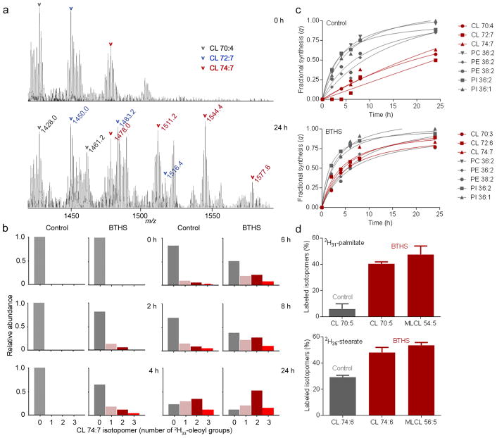
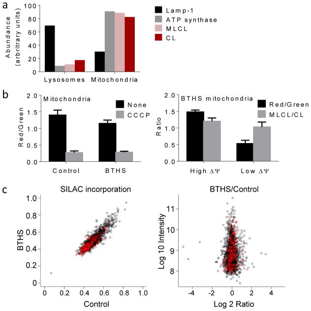
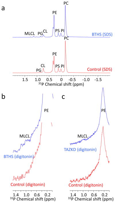
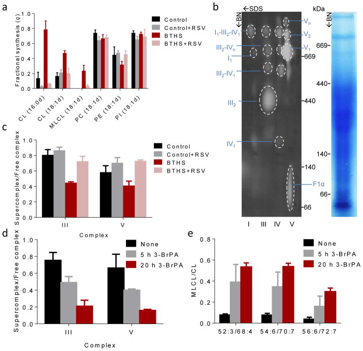
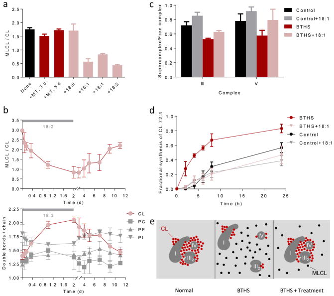
Cardiolipin is a specific mitochondrial phospholipid that has a high affinity for proteins and that stabilizes the assembly of supercomplexes involved in oxidative phosphorylation. We found that sequestration of cardiolipin in protein complexes is critical to protect it from degradation. The turnover of cardiolipin is slower by almost an order of magnitude than the turnover of other phospholipids. However, in subjects with Barth syndrome, cardiolipin is rapidly degraded via the intermediate monolyso-cardiolipin. Treatments that induce supercomplex assembly decrease the turnover of cardiolipin and the concentration of monolyso-cardiolipin, whereas dissociation of supercomplexes has the opposite effect. Our data suggest that cardiolipin is uniquely protected from normal lipid turnover by its association with proteins, but this association is compromised in subjects with Barth syndrome, leading cardiolipin to become unstable, which in turn causes the accumulation of monolyso-cardiolipin.


Conflict of interest statement

COMPETING FINANCIAL INTERESTS

The authors declare no competing financial interests.

Figures

Figure 1
Figure 1. BTHS increases the acyl turnover of CL in human lymphoblasts
(a) Human lymphoblasts were incubated with 2H33-oleic acid; lipids were analyzed by MS. The CL region of the raw spectra is shown before (0 h) and after (24 h) labeling. Novel isotopomers formed as a result of 2H33-oleoyl incorporation. (b) Intensities of CL 74:7 isotopomers with 0, 1, 2, and 3 2H33-oleoyl groups were determined at different time points. The isotopomer pattern changed faster in BTHS lymphoblasts than in controls. Data are means of duplicate determinations. (c) The fractional syntheses of the indicated lipid species were determined at different time points. The turnover of CL was slower than the turnover of other phospholipids in controls but not in BTHS. Statistical analysis of the data is shown in Supplementary Table 1. (d) Human lymphoblasts were incubated with 2H31-palmitic acid or 2H35-stearic acid for 20 hours. Lipids were analyzed by MS to determine the proportion of labeled isotopomers [labeled/(labeled+unlabeled)]. BTHS caused increased labeling of CL with 2H31-palmitate (P<0.0001) and 2H35-stearate (P<0.002). The proportion of labeled isotopomers was similar in CL and MLCL; the latter was present only in BTHS. Data (means±s.e.m., N=4) were analyzed by t-test.
Figure 2
Figure 2. Tafazzin deficiency increases the glycerol turnover of CL in Drosophila and mammalian cells
(a) Drosophila strains were cultured for 9 days with D-[U-13C6]-glucose or unlabeled glucose as control. Lipids were analyzed by MS and spectra are displayed in centroid mode. 13C was incorporated into PE of the wild-type (WT) and the tafazzin deletion strain (ΔTAZ). 13C was incorporated into CL and MLCL of ΔTAZ but not WT, indicating a very slow CL turnover in the WT. (b) The fractional syntheses of CL and MLCL species were determined from the relative abundances of 13C0, 13C3,13C6 and 13C9 isotopomers following labeling with D-[U-13C6]-glucose for the indicated time periods. Tafazzin deficiency caused an increase in the fractional synthesis of CL species in Drosophila (P<0.04, 3 d; P<0.01, 9 d), mouse fibroblasts (P<0.001), and human lymphoblasts (P<0.0001). The fractional syntheses of CL and MLCL were similar. Data (means±s.e.m., N=3) were analyzed by t-test.
Figure 3
Figure 3. MLCL is present in intact mitochondria with normal protein turnover
(a) Lysosomes and mitochondria were prepared from BTHS lymphoblasts. Marker proteins for lysosomes (Lamp-1) and mitochondria (α-subunit of ATP synthase) were quantified by Western blotting; MLCL and CL were quantified by MS. CL and MLCL were associated with mitochondria. Data are means of duplicate determinations. (b) Mitochondria were isolated from human lymphoblasts and analyzed by flow cytometry after staining with JC-1. The membrane potential, measured by the red/green fluorescence ratio, was slightly lower in BTHS mitochondria than in controls (P<0.03, t-test). CCCP collapsed the membrane potential. Isolated BTHS mitochondria were divided with a cell sorter into two populations one with high and one with low membrane potential (ΔΨ). The populations were analyzed by flow cytometry and MS, which confirmed the difference in membrane potential but did not show any difference in the MLCL/CL ratio. Data are means±s.e.m. (N=3). (c) Human lymphoblasts were cultured with stable isotope labeled amino acids. The level of incorporation of labeled amino acids into mitochondrial (red circles) and non-mitochondrial (black circles) proteins was measured by proteomic analysis. The BTHS/control ratio of incorporated label was measured in 1:1 mixtures of control and BTHS samples, each labeled with different precursors. The turnover of mitochondrial proteins was similar in BTHS and controls.
Figure 4
Figure 4. CL but not MLCL is associated with proteins
(a) Mitochondrial membranes were prepared from human lymphoblasts and solubilized with SDS. 31P-NMR spectra show resonances of all major and some minor phospholipids. CL was detectable in control samples; CL and MLCL were detectable in BTHS samples. (b–c) Mitochondrial membranes, prepared from human lymphoblasts (b) or mouse kidneys (c), were solubilized with digitonin. 31P-NMR spectra show the major phospholipids with broader resonances compared to the SDS experiment. MLCL (chemical shift values of MLCL standard: 1.0, 1.2 ppm) was detectable in tafazzin-deficient samples but CL was not.
Figure 5
Figure 5. Supercomplexes protect CL from degradation
(a) Human lymphoblasts were cultured ±40 μM RSV for 36 hours. The fractional syntheses of lipids were determined by incorporating 2H-labeled palmitic acid (16:0d) or oleic acid (18:1d) for 8 hours. RSV inhibited the turnover of CL in BTHS (P<0.03) but not in controls. RSV had no effect on other phospholipids. (bc) Lymphoblast mitochondria were solubilized with digitonin, separated by 2D-Blue Native/SDS-PAGE, and analyzed with primary antibodies to NDUFB6 (complex I), UQCRC2 (complex III), MtCO1 (complex IV), and F1α (complex V). The Coomassie-stained lane shows the BN-PAGE separation and the 2-D blot shows individual complexes and supercomplexes with the positions of molecular weight markers. BTHS decreased the proportion of supercomplexes, RSV increased it back to normal (P<0.02). (de) Control lymphoblasts were treated with 80 μM 3-bromopyruvate (3-BrPA) for 5 or 20 hours. 3-BrPA decreased the proportion of supercomplexes and increased the MLCL/CL ratio (P<0.03). Data (means±s.e.m., N=3) were analyzed by t-test (a, c) or ANOVA (d, e).
Figure 6
Figure 6. Unsaturated fatty acids stabilize CL
(a) BTHS lymphoblasts were cultured with 5 μM MitoTempo (MT) for 3 or 5 days or with 0.1 mM exogenous fatty acid (stearic acid, 18:0; palmitoleic acid, 16:1; oleic acid, 18:1; linoleic acid, 18:2) for 4 days. Unsaturated fatty acids decreased the MLCL/CL ratio (P<0.003, t-test). (b) BTHS lymphoblasts were cultured first with and then without linoleic acid. The MLCL/CL ratio inversely correlated with the CL unsaturation. (c–d) Lymphoblasts were cultured ± 18:1 for 4 days followed by measurements of supercomplex abundance and CL turnover with 2H33-oleic acid. BTHS reduced the proportion of supercomplexes and increased the turnover of CL (P<0.03, t-test). 18:1 increased the proportion of supercomplexes and reduced the turnover of CL in BTHS but not in controls (P<0.02, t-test). Half-lives of CL 72:4, estimated by non-linear regression analysis, were 20 hours (control), 30 hours (control+18:1), 5 hours (BTHS), and 26 hours (BTHS+18:1). Data in panels a–d are means±s.e.m. (N=3). (e) The cartoon shows supercomplex I1-III2-IV1 as an example; a similar mechanism is suggested for other complexes. In normal mitochondria, unsaturated CL clusters around protein complexes, which promotes their assembly to supercomplexes and protects CL from degradation. In BTHS, the fatty acid composition of CL is altered. Less CL is associated with proteins and non-associated CL is hydrolyzed to MLCL. Treatments that increase CL unsaturation or force the formation of supercomplexes, increase the proportion of protein-associated CL. As a result, less CL is hydrolyzed to MLCL.

References

    1. Mileykovskaya E, Dowhan W. Cardiolipin-dependent formation of mitochondrial respiratory supercomplexes. Chem Phys Lipids. 2014;179:42–48. - PMC - PubMed
    1. Ren M, Phoon CKL, Schlame M. Metabolism and function of mitochondrial cardiolipin. Progr Lipid Res. 2014;55:1–16. - PubMed
    1. Lewis RNAH, McElhaney RN. The physicochemical properties of cardiolipin bilayers and cardiolipin-containing lipid membranes. Biochim Biophys Acta. 2009;1788:2069–2079. - PubMed
    1. Zhang M, Mileykovskaya E, Dowhan W. Gluing the respiratory chain together. Cardiolipin is required for supercomplex formation in the inner mitochondrial membrane. J Biol Chem. 2002;277:43553–43556. - PubMed
    1. Althoff T, Mills DJ, Popot JL, Kühlbrandt W. Arrangement of electron transport chain components in bovine mitochondrial supercomplex I1III2IV1. EMBO J. 2011;30:4652–4664. - PMC - PubMed

Publication types

LinkOut - more resources