. 2016 Aug 1;126(8):2821-6.
doi: 10.1172/JCI84705. Epub 2016 Jun 27.

Pneumococcal meningitis is promoted by single cocci expressing pilus adhesin RrgA

Pneumococcal meningitis is promoted by single cocci expressing pilus adhesin RrgA

Federico Iovino et al. J Clin Invest. .

Abstract

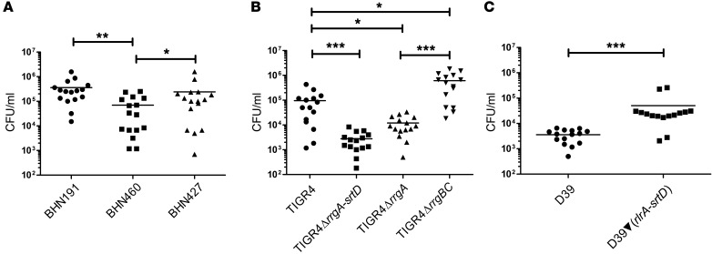
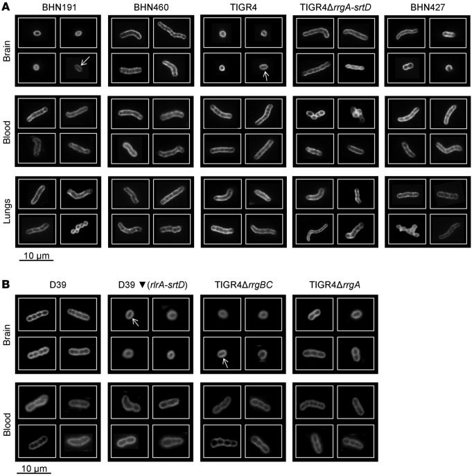
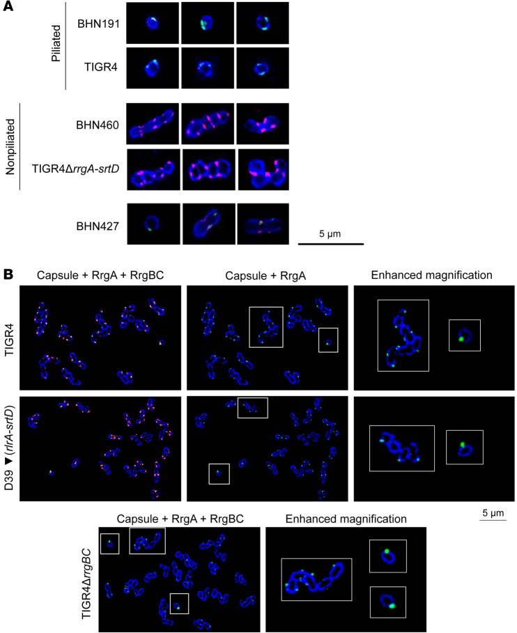
Streptococcus pneumoniae (pneumococcus) is the primary cause of bacterial meningitis. Pneumococcal bacteria penetrates the blood-brain barrier (BBB), but the bacterial factors that enable this process are not known. Here, we determined that expression of pneumococcal pilus-1, which includes the pilus adhesin RrgA, promotes bacterial penetration through the BBB in a mouse model. S. pneumoniae that colonized the respiratory epithelium and grew in the bloodstream were chains of variable lengths; however, the pneumococci that entered the brain were division-competent, spherical, single cocci that expressed adhesive RrgA-containing pili. The cell division protein DivIVA, which is required for an ovoid shape, was localized at the poles and septum of pneumococcal chains of ovoid, nonseparated bacteria, but was absent in spherical, single cocci. In the bloodstream, a small percentage of pneumococci appeared as piliated, RrgA-expressing, DivIVA-negative single cocci, suggesting that only a minority of S. pneumoniae are poised to cross the BBB. Together, our data indicate that small bacterial cell size, which is signified by the absence of DivIVA, and the presence of an adhesive RrgA-containing pilus-1 mediate pneumococcal passage from the bloodstream through the BBB into the brain to cause lethal meningitis.


Figures

Figure 1
Figure 1. Pilus-1 promotes invasion of S. pneumoniae into the brain.
C57BL/6 mice were infected i.v. with (A) serotype 6B isolates, (B) TIGR4 and isogenic pilus mutants, and (C) D39 and piliated D39▼(rlrA-srtD). Graphs show the number of pneumococci found in the brain: each dot represents 1 mouse; black bars show the average values. n = 15 per group (3 experiments with n = 5 per group). *P < 0.05, **P < 0.01, and ***P < 0.001, by nonparametric ANOVA, followed by Dunn’s test (A and B) and by nonparametric, 2-tailed Wilcoxon’s rank-sum test (C).
Figure 2
Figure 2. Pneumococci carrying pilus-1 invade the brain as single cocci.
(A) Immunofluorescence staining of the polysaccharide capsule of 6B clinical isolates, TIGR4, and TIGR4ΔrrgA-srtD from brain and lung homogenates and blood and (B) D39, D39▼(rlrA-srtD), TIGR4ΔrrgBC, and TIGR4ΔrrgA from brain homogenates and blood. A mixed phenotype was found for BHN427 (A), and in the brain, among piliated strains, only a minor part formed diplococci (see detailed quantification in the Supplemental Information). For each strain and each type of tissue, 3 mice were analyzed (approximately 300 images per mouse were taken).
Figure 3
Figure 3. In the blood, piliated and RrgA-expressing single cocci, typified by the absence of DivIVA, express more RrgA than do chains and are more prone to penetration of the BBB.
(A) Immunofluorescence staining showing capsule (blue), pilus-1 (green), and DivIVA (red) of pneumococci in brain homogenates. For each strain, 3 mice were analyzed (approximately 300 images per mouse were taken). (B) Immunofluorescence staining of RrgA-expressing pneumococci in blood samples showing capsule (blue), RrgA (green), and RrgBC (red). “Enhanced magnification” panels show the parts within the white-outlined areas at higher magnification (original magnification, ×5). One representative image per strain is shown (×100 objective); in total, approximately 900 bacteria per strain were imaged.

References

    1. Brouwer MC, Tunkel AR, van de Beek D. Epidemiology, diagnosis, and antimicrobial treatment of acute bacterial meningitis. Clin Microbiol Rev. 2010;23(3):467–492. doi: 10.1128/CMR.00070-09. - DOI - PMC - PubMed
    1. Browall S, et al. Intraclonal variations among Streptococcus pneumoniae isolates influence the likelihood of invasive disease in children. J Infect Dis. 2014;209(3):377–388. doi: 10.1093/infdis/jit481. - DOI - PMC - PubMed
    1. Weiser JN. The battle with the host over microbial size. Curr Opin Microbiol. 2013;16(1):59–62. doi: 10.1016/j.mib.2013.01.001. - DOI - PMC - PubMed
    1. Mook-Kanamori BB, Geldhoff M, van der Poll T, van de Beek D. Pathogenesis and pathophysiology of pneumococcal meningitis. Clin Microbiol Rev. 2011;24(3):557–591. doi: 10.1128/CMR.00008-11. - DOI - PMC - PubMed
    1. Orihuela CJ, et al. Laminin receptor initiates bacterial contact with the blood brain barrier in experimental meningitis models. J Clin Invest. 2009;119(6):1638–1646. doi: 10.1172/JCI36759. - DOI - PMC - PubMed

Publication types