. 2016 Jun 28:7:11963.
doi: 10.1038/ncomms11963.

Memory of cell shape biases stochastic fate decision-making despite mitotic rounding

Affiliations

Memory of cell shape biases stochastic fate decision-making despite mitotic rounding

Takashi Akanuma et al. Nat Commun. .

Abstract

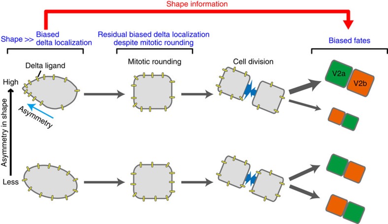
Cell shape influences function, and the current model suggests that such shape effect is transient. However, cells dynamically change their shapes, thus, the critical question is whether shape information remains influential on future cell function even after the original shape is lost. We address this question by integrating experimental and computational approaches. Quantitative live imaging of asymmetric cell-fate decision-making and their live shape manipulation demonstrates that cellular eccentricity of progenitor cell indeed biases stochastic fate decisions of daughter cells despite mitotic rounding. Modelling and simulation indicates that polarized localization of Delta protein instructs by the progenitor eccentricity is an origin of the bias. Simulation with varying parameters predicts that diffusion rate and abundance of Delta molecules quantitatively influence the bias. These predictions are experimentally validated by physical and genetic methods, showing that cells exploit a mechanism reported herein to influence their future fates based on their past shape despite dynamic shape changes.


Figures

Figure 1
Figure 1. Quantitative definition of cellular eccentricity of V2 cells and its relation to their fates.
(a) Dynamics of shape changes of V2 cell. (b) Quantitative representation of the direction and degree of the asymmetric elongation of V2 cell shape. (c) Typical examples of less (top) and highly (bottom) asymmetric cell shapes in 3D. (d) The distribution of various Along values of all V2 cells (n=314) analysed in this study. See also Supplementary Fig. 1 and Supplementary Movies 1 and 2. (e) Orientating (+ or −) the positions of the two daughter cells (V2a and V2b) by fate axis. (f) Bias in the stochastic fate decision-making according to Along values. Receiver-operating characteristic analysis found the threshold for the shape-fate relation is Along=0.036. Red dotted line indicates 50% incidence. Scale bar, 10 μm. NS, not significant. *P<0.05 (χ2-test).
Figure 2
Figure 2. V2 cellular eccentricity biases stochastic fate decision-making.
(a) Alteration of the direction of the asymmetric elongation of the V2 cell shape by femtosecond laser irradiation. The spiky ends of the old (yellow arrow) and new (orange arrow) axes are indicated. (b) Along values, axis changes and θfate for individual V2 cells on femtosecond laser irradiation are shown as 3D plot. The two-dimensional plots of axis change—Along and axis change—θfate are shown in c and d, respectively. Plotted are the cells with very little axis change (<45°) and θfate >90° (red open circle), significant axis change (>45°) and θfate >90° (blue open circle), little axis change (<45°) and θfate <90° (red closed circle) and significant axis change (>45°) and θfate <90° (blue closed circle). (e) Fates of the laser-irradiated V2 cells. (f) Summary. Fate decisions are made according to the newly acquired orientation of V2 cell shape asymmetry. Scale bar, 10 μm. NS, not significant. *P<0.05, **P<0.01 (χ2-test).
Figure 3
Figure 3. Model for shape-dependent biased fate decision-making and in silico validation of the model by computer simulations.
(a) The lateral inhibition system generated by the direct Delta–Notch trans-engagement at the contacting cell surface. (b) A mechanistic model for the shape-dependent biased fate decision-making. (c) Force-generating system to drive cell division. (d) Integration of both cell division and fate decision-making systems into one model based on 3D CPM. (e) Computer simulation of the shape-dependent biased fate decision-making model. Simulation results with in silico cells of three relatively symmetrical (Along=0.002, 0.024 and 0.031) and three asymmetrical (Along=0.052, 0.063 and 0.092) shapes are shown. NS, not significant. *P<0.05, **P<0.01 (χ2-test). See also Supplementary Fig. 3 and Supplementary Movies 3 and 4.
Figure 4
Figure 4. Simulating the effects of Delta molecule dynamics on the bias.
(a) No bias in the fate decision-making with no initial polarized localization of Delta particles, despite the asymmetrical shape. Simulation results with three asymmetrical (Along=0.052, 0.063 and 0.092) shapes are shown. (b) Diffusion rate-dependent bias in the fate decision-making of the in silico cells of asymmetrical shapes. Higher diffusion rate: four lattice site length per mcs. Simulation results with three asymmetrical (Along=0.052, 0.063 and 0.092) shapes are shown. (c) Weaker and stronger biases in the fate decision-making with fewer (0.0025 particle concentration (conc.)/diffusion rate) or more Delta particles (0.25 particle conc./diffusion rate), respectively. Simulation results with three asymmetrical (Along=0.052, 0.063 and 0.092) shapes are shown. NS, not significant, *P<0.05; **P<0.01 (χ2-test).
Figure 5
Figure 5. V2 cell eccentricity polarizes the localization of DeltaC protein.
(a) Quantitative measurement of DeltaC protein distribution. (b) Polarized localization of DeltaC protein on the (+) side of V2 cell. (c) Diffusion-like behaviour of the initially polarized DeltaC::mCherry fusion protein during mitotic cell rounding. (d) Translocation of DeltaC::mCherry fusion protein on changes of the direction of the asymmetric elongation of V2 cell shape. The spiky ends of the old (yellow arrow) and new (orange arrow) axes are indicated. Irradiation at the spiky ends of the old axis (yellow arrow) altered the asymmetry orientation generating the new axis. Biased localization of the DeltaC::mCherry fusion protein is shifted according to the newly acquired orientation of V2 cell shape asymmetry. *P<0.05, **P<0.01 (χ2-test). Scale bars, 5 μm (b), 10 μm (c,d). See also Supplementary Fig. 4 and Supplementary Movies 5 and 6.
Figure 6
Figure 6. DeltaC is an essential mediator of the ‘shape-memory' system.
(a) Failure of V2a/V2b fate decision-making in DeltaC-deficient V2 cell and its rescue by DeltaC re-expression. (b) Reduced Notch-signalling activity in DeltaC-deficient V2 cell and its rescue by DeltaC re-expression. See Methods for the details of this assay system and the data analyses. (c) Rescue of the biased fate decision-making accompanied by the biased DeltaC protein localization in the deltaC-deficient V2 cells by re-expressing DeltaC. The spiky ends of the old (yellow arrow) and new (orange arrow) axes are indicated. *P<0.05 (χ2-test). Scale bars, 10 μm.
Figure 7
Figure 7. The bias is dependent on the abundance of DeltaC molecules.
(a) Altered DeltaC protein levels by loss- and gain-of-functions. Scale bar, 10 μm. (b) Attenuation of the bias by the reduced DeltaC protein level. (c) Enhancement of the bias by the increased DeltaC protein level. NS, not significant. **P<0.01 (χ2-test). See also Supplementary Fig. 5.
Figure 8
Figure 8. Schematic diagram of the ‘shape-memory' system medicated by the biased DeltaC protein localization.
Polarized DeltaC localization caused by V2 cell eccentricity biases its stochastic fate decision-making even after mitotic rounding.

Comment in

  • Shaping the cell fate.
    Sato TN, Merks RM. Sato TN, et al. Cell Cycle. 2017 Jan 17;16(2):149-150. doi: 10.1080/15384101.2016.1241603. Epub 2016 Oct 13. Cell Cycle. 2017. PMID: 27736341 Free PMC article. No abstract available.

Similar articles

Cited by

References

    1. Hertwig O. Das Problem der Fefrunchtung une der isotropie des Eies, eine Theory der Vererbung. Jenaische Zeitschrift für Naturwissenschaft 18, 276–318 (1884).
    1. Minc N., Burgess D. & Chang F. Influence of cell geometry on division-plane positioning. Cell 144, 414–426 (2011). - PMC - PubMed
    1. Minc N. & Piel M. Predicting division plane position and orientation. Trends Cell Biol. 22, 193–200 (2012). - PubMed
    1. Folkman J. & Moscona A. Role of cell shape in growth control. Nature 273, 345–349 (1978). - PubMed
    1. Meyers J., Craig J. & Odde D. J. Potential for control of signaling pathways via cell size and shape. Curr. Biol. 16, 1685–1693 (2006). - PubMed

Publication types

Substances

LinkOut - more resources