. 2016 Jun 28:6:28686.
doi: 10.1038/srep28686.

Functional map of arrestin binding to phosphorylated opsin, with and without agonist

Affiliations

Functional map of arrestin binding to phosphorylated opsin, with and without agonist

Christian Peterhans et al. Sci Rep. .

Erratum in

Abstract

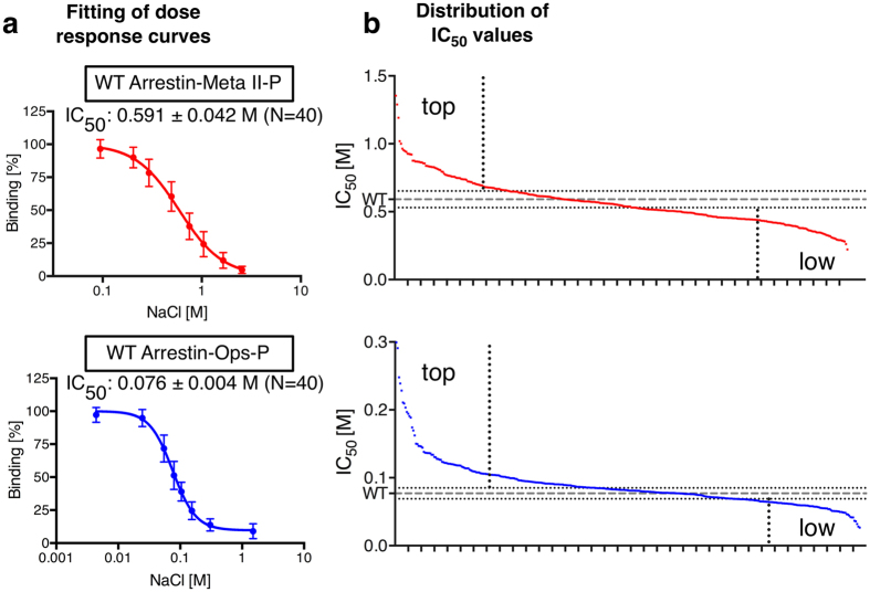
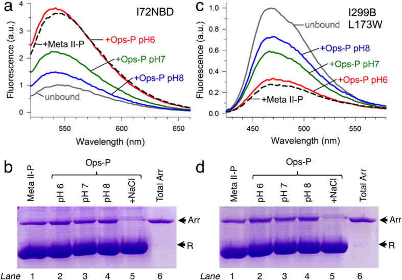
Arrestins desensitize G protein-coupled receptors (GPCRs) and act as mediators of signalling. Here we investigated the interactions of arrestin-1 with two functionally distinct forms of the dim-light photoreceptor rhodopsin. Using unbiased scanning mutagenesis we probed the individual contribution of each arrestin residue to the interaction with the phosphorylated apo-receptor (Ops-P) and the agonist-bound form (Meta II-P). Disruption of the polar core or displacement of the C-tail strengthened binding to both receptor forms. In contrast, mutations of phosphate-binding residues (phosphosensors) suggest the phosphorylated receptor C-terminus binds arrestin differently for Meta II-P and Ops-P. Likewise, mutations within the inter-domain interface, variations in the receptor-binding loops and the C-edge of arrestin reveal different binding modes. In summary, our results indicate that arrestin-1 binding to Meta II-P and Ops-P is similarly dependent on arrestin activation, although the complexes formed with these two receptor forms are structurally distinct.


Figures

Figure 1
Figure 1. Quantification of arrestin mutants and calculation of IC50 values.
Each alanine-mutant was expressed in E. coli and used for centrifugal pull-down analysis with native ROS membranes containing Meta II-P or Ops-P. Light-activated Meta II-P was assayed in parallel and simultaneously with the apo-form Ops-P. (a) NaCl titrations against arrestin-1 wild-type (WT) binding to Meta II-P (red) or Ops-P (blue) were used to calculate sigmoidal dose-response curves and extract IC50 values. (b) Distribution of IC50 values for the complete arrestin functional map when bound to phosphorylated Meta II-P (red) and Ops-P (blue). The dashed line indicates the WT IC50 values, and dotted lines indicate the doubled standard deviation calculated from arrestin WT data sets.
Figure 2
Figure 2. Functional test of receptor phosphorylation.
The “Extra Meta II” assay measures the stabilisation of Meta II (λmax: 380 nm) over its precursor Meta I (λmax: 480 nm) by absorbance spectroscopy. In the absence of any binding partner, very little Meta II evolved (black trace). In the presence of the high-affinity analogue peptide derived from the C-terminus of the α-subunit of transducin (Gtα-peptide, VLEDLKSCGLF, 350 μM), 100% of receptors were stabilised as Meta II (blue trace). In the presence of arrestin (10 μM), all receptors were also stabilised as Meta II (red trace). Hence, all receptors in our preparation were sufficiently phosphorylated to bind arrestin as Meta II-P. A high level of receptor phosphorylation is indicated by the fast rate of arrestin binding, which is comparable to Gtα-peptide. This assay has previously been established as an indicator of functional receptor phosphorylation for studies of arrestin binding.
Figure 3
Figure 3. Functional maps of arrestin-1 binding to Meta II-P and Ops-P.
(a) Important functional regions highlighted on the basal arrestin structure. (b) IC50 values for Meta II-P binding plotted on the basal structure of arrestin-1. Mutations which significantly increased or decreased binding are coloured green and purple, respectively. (c) IC50 values for Ops-P binding. Colour code follows that described in (a). (d) Positive correlation of mutants for Meta II-P and Ops-P binding. Green – mutations which increased binding for both Meta II-P and Ops-P. Orange – mutations which decreased binding for both Meta II-P and Ops-P. (e) Negative correlation of mutants for Meta II-P and Ops-P binding. Yellow – mutations which increased affinity for Meta II-P but decreased affinity for Ops-P. Pink – mutations which decreased affinity for Meta II-P but increased affinity for Ops-P. For (be), only IC50 values outside the doubled standard deviation are shown.
Figure 4
Figure 4. Site-directed fluorescence.
(a) Steady-state fluorescence spectra of arrestin I72NBD (1 μM) were measured in the absence (grey trace) or presence of an excess of receptor (6 μM): light-activated Meta II-P at pH 7 (dashed black trace), Ops-P at pH 6 (red trace), Ops-P at pH 7 (green trace) or OpsP at pH 8 (blue trace). (b) Centrifugal pull-down analysis of arrestin I72NBD binding to Meta II-P at pH 7 (lane 1) or Ops-P at pH 6, 7 or 8 (lanes 2, 3 and 4, respectively). As a negative control, no pull down of arrestin with Ops-P at pH 7 in the presence of 1 M NaCl was observed (lane 5). The total amount of arrestin in each pull-down experiment (4.5 μg) is shown in lane 6. The arrows indicate the location of arrestin (Arr) and receptor (R). (c) Same as described in (a), performed with arrestin I299B/L173W. (d) Same as described in (b), performed with arrestin I299B/L173W. Note that these experiments were performed in low-salt buffer in order to maximize arrestin binding to OpsP.
Figure 5
Figure 5. Different engagement of phosphosensors.
Phosphosensing residues, whose mutation strongly decreased binding are highlighted in blue in (a) and (b) on pre-activated arrestin p44. (a) Phosphosensitive residues deployed by Meta II-P (K14, K15, K20, R29, K110, K300 and H301) are located along the side of the arrestin N-domain. Side view (left) and top view (right) of arrestin are shown. The top view represents the orientation of arrestin as seen from the cytoplasmic face of the receptor. (b) Phosphosensing residues deployed by Ops-P (K14, K15, R18, K55, R56, R81, K150, K166, R171 and K300) are located in the cup of the arrestin N-domain. Views of arrestin are as in (a). (c) The phosphosensitive residues (blue) determined from the Meta II-P functional map are plotted on the Ops*/arrestin-1 complex. The receptor is cyan, and the dashed line indicates the membrane plane. (d) The receptor phosphopeptide (Rpp) (mint green) aligned on arrestin. The Rpp model is adapted from. N326, shown as sticks, is the last resolved residue in and is also shown as sticks in the Rpp model.
Figure 6
Figure 6. Mutational data on the Ops*/arrestin-1 complex structure.
(a) Model derived from the arrestin-receptor complex structure, rhodopsin (cyan), and arrestin (grey). Finger, middle and C-loop are coloured like in Fig. 3. Dashed lines indicate the membrane bilayer. (b) Arrestin-rhodopsin interaction coloured by IC50 values derived by Meta II-P interaction. (c) Arrestin-rhodopsin interaction coloured by IC50 values derived by Ops-P interaction. In (b,c), the receptor-arrestin interaction, coloured according to mutations which significantly increased (green) or decreased (purple) binding for Ops-P (colour code follows that from Fig. 3b,c). Core interaction residues as classified by EPPIC are shown as sticks. Top panels in (b,c) show the interaction with loop 160, finger, middle and C-loop. Bottom panels show putative membrane interaction sites in the C-edge loops.
Figure 7
Figure 7. Interaction of arrestin with different functional forms of the receptor.
(a) Cartoon representation of arrestin in the basal state. Notable loops include the finger loop (blue), the gate loop (yellow) and the 160-loop (black). The C-tail of arrestin is dark orange and interacts with the N-domain through the 3-element interaction and the polar core. (b) Dark state rhodopsin (Rho, red) and basal arrestin. (c) Light activation converts rhodopsin to Meta II, which is phosphorylated on the C-terminus by GRK1 and then bound by arrestin. Hallmarks of high affinity binding include receptor engagement of the finger loop and 160-loop, inter-domain rotation, and movement of the gate loop, which allows the phosphorylated receptor C-terminus to bind within a positively charged cleft in the N-domain. (d,e) Meta II-P decays to the apo-receptor Ops-P, which exists in a conformational equilibrium between an active (Ops*) and inactive (Ops) form. Arrestin binds Ops*-P similarly as Meta II-P. The interaction of arrestin with inactive Ops-P differs in the placement of the finger loop, the phosphorylated receptor C-terminus and how the C-edge is engaged.

Similar articles

Cited by

References

    1. Fredriksson R., Lagerström M. C., Lundin L.-G. & Schiöth H. B. The G-protein-coupled receptors in the human genome form five main families. Phylogenetic analysis, paralogon groups, and fingerprints. Mol. Pharmacol. 63, 1256–1272 (2003). - PubMed
    1. Lefkowitz R. J. Seven transmembrane receptors: something old, something new. Acta Physiol (Oxf) 190, 9–19 (2007). - PubMed
    1. Schöneberg T. T. et al.. Mutant G-protein-coupled receptors as a cause of human diseases. Pharmacol. Ther. 104, 173–206 (2004). - PubMed
    1. Gurevich E. V. & Gurevich V. V. Arrestins: ubiquitous regulators of cellular signaling pathways. Genome Biol. 7, 236 (2006). - PMC - PubMed
    1. Nobles K. N. et al.. Distinct phosphorylation sites on the β(2)-adrenergic receptor establish a barcode that encodes differential functions of β-arrestin. Sci Signal 4, ra51 (2011). - PMC - PubMed

Publication types