. 2017 Jan;35(1):170-180.
doi: 10.1002/stem.2451. Epub 2016 Jul 21.

Angiopellosis as an Alternative Mechanism of Cell Extravasation

Affiliations

Angiopellosis as an Alternative Mechanism of Cell Extravasation

Tyler A Allen et al. Stem Cells. 2017 Jan.

Abstract

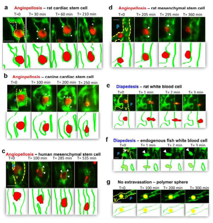
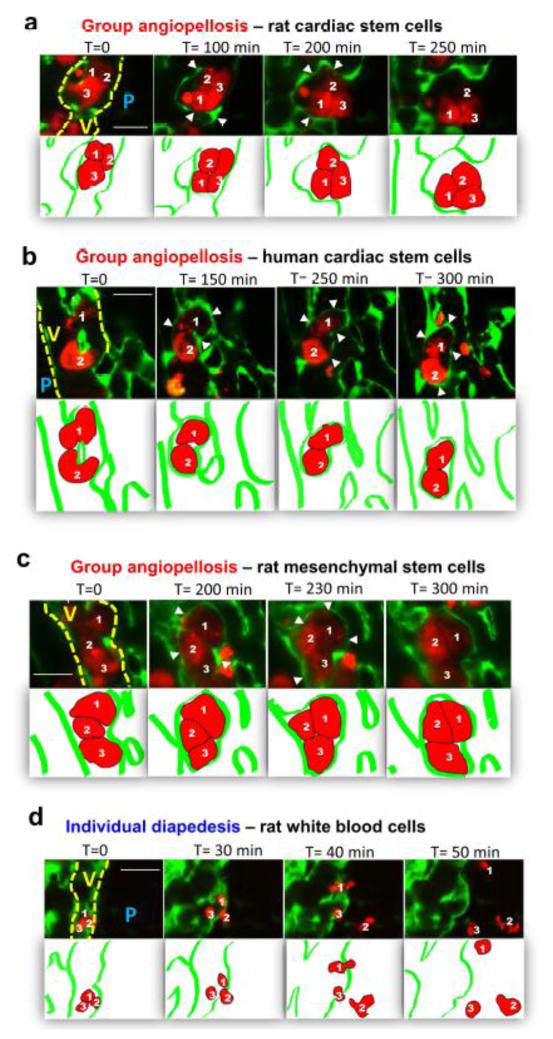
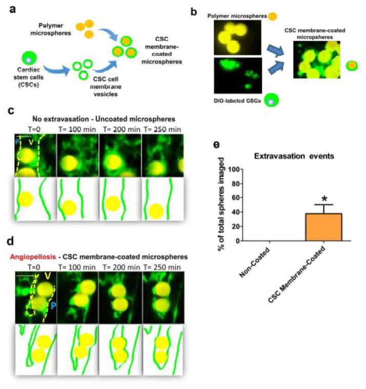
Stem cells possess the ability to home in and travel to damaged tissue when injected intravenously. For the cells to exert their therapeutic effect, they must cross the blood vessel wall and enter the surrounding tissues. The mechanism of extravasation injected stem cells employ for exit has yet to be characterized. Using intravital microscopy and a transgenic zebrafish line Tg(fli1a:egpf) with GFP-expressing vasculature, we documented the detailed extravasation processes in vivo for injected stem cells in comparison to white blood cells (WBCs). While WBCs left the blood vessels by the standard diapedesis process, injected cardiac and mesenchymal stem cells underwent a distinct method of extravasation that was markedly different from diapedesis. Here, the vascular wall undergoes an extensive remodeling to allow the cell to exit the lumen, while the injected cell remains distinctively passive in activity. We termed this process Angio-pello-sis, which represents an alternative mechanism of cell extravasation to the prevailing theory of diapedesis. Stem Cells 2017;35:170-180 Video Highlight: https://youtu.be/i5EI-ZvhBps.

Keywords: Angiopellosis; Diapedesis; Extravasation; Stem cell infusion; Transmigration.


Conflict of interest statement

Competing Financial Interests

The authors declare no competing financial interests.

Figures

Figure 1
Figure 1. Intravital imaging of zebrafish vasculature using light-sheet microscopy for visualization of cell extravasation
(a) Schematic illustration of angiopellosis in which endothelial cells extend protrusions around injected stem cells and actively expulse injected cell(s) into the parenchyma. Green cells represent green fluorescent protein (GFP)-expressing endothelial cells of the transgenic Tg(fli1a:EGFP) zebrafish embryo blood vessels; red cell represents injected stem cells; white arrow indicated blood flow. (b) Schematic illustration of cell injection in Tg(fli1a:EGFP) zebrafish embryos. (b-α) Region of injection (red box) and injected cells (arrow) in the cardiac region following the duct of cuvier of a 48 hpf zebrafish embryo; (b-β) Region of imaging in the tail-area vessels (TAVs) zone (red box). (c) Schematic showing the set-up of light-sheet microscopy. The living zebrafish is embedded in 1.3% agarose gel and positioned in front of the water-dipping detection lens. The sheet of light is generated by fast vertical scanning of a focused laser beam, and it illuminates a 4-μm-thick volume section of the fish. Fluorescence is recorded orthogonally to the light sheet with a wide-field detection arm equipped with a fast scientific complementary metal-oxide semiconductor camera. Fast volumetric imaging is performed by step-wise axial movement of the detection objective in synchrony with displacement of the light sheet while the specimen is kept stationary. (d) Three-dimensional rendering of z-stack images of the tail-area vessels (green) with injected cells (red). (e) Tail-area vessel (TAV) region of a double transgenic zebrafish Tg(fli1a:EGFP)/(mpeg1:EGFP) embryo, in which GFP is expressed simultaneously in vasculature and macrophages. Arrows indicate macrophages expressing GFP. Scale bar = 50 μm.
Figure 2
Figure 2. Injected cardiac stem cells undergo extravasation by angiopellosis
(a) Time-lapse imaging in Tg(fli1a:EGFP) zebrafish embryos shows intravenously injected rat cardiac stem cell (red) after becoming lodged in the blood vessel (green); T=time in minutes. Gradual protrusions of the vascular endothelial cells can be seen remodeling around the injected stem cell (T=30, arrows). By 3.5 hour the stem cell has completely undergone angiopellosis, and is fully extravasated (T=210). Residual remodeling from the endothelial protrusions are still present and active up until 210 minutes after extravasation (T=60, T=210, arrows). (b) Time-lapse imaging of canine cardiac stem cells (red) extravasating blood vessel (green). Vascular remodeling (T=100, arrows) can be seen actively interacting with the stem cell. Injected stem cell fully extravasates after approximately 250 minutes (T=250). (c) Time-lapse imaging of human mesenchymal stem cells (red) extravasating blood vessel (green). Vascular remodeling (T=100, arrows) can be seen actively interacting with the stem cell. Injected stem cell fully extravasates after approximately 535 minutes (T=535) (d) Time-lapse imaging of human rat stem cells (red) extravasating blood vessel (green). Vascular remodeling (T=205, arrows) can be seen actively interacting with the stem cell. Injected stem cell fully extravasates after approximately 6 hours (T=360). (e) Time-lapse imaging in Tg(fli1a:EGFP) zebrafish embryos shows intravenously injected rat white blood cell (red) undergoing diapedesis to extravasate out the zebrafish blood vessel (green). (f) Time-lapse imaging in double transgenic Tg(fli1a:EGFP)/ (mpeg1:EGFP), in which both vasculature and endogenous macrophages express GFP. Zebrafish embryos endogenous white blood cell (arrow) undergo diapedesis to extravasate blood vessel in characteristic manner. (g) Time-lapse imaging in Tg(fli1a:EGFP) zebrafish embryos shows intravenously inert 8 μm polymer microspheres (yellow), the microsphere remains in the relatively same position over the course of approximately 300 minutes. Yellow dotted line represents the vascular wall of the blood vessel. V=vasculature lumen; P= parenchymal surrounding tissue. All scale bars = 20 μm.
Figure 3
Figure 3. Multiple CSCs undergo cluster extravasation in a single angiopellosis event
(a) Time-lapse imaging in Tg(fli1a:EGFP) zebrafish embryos shows intravenously (IV) injected rat cardiac stem cell (red) cluster lodged in the blood vessel (green); T=time in minutes. Clustered cells underwent extravasation with endothelial protrusions (arrows) extending around three injected cells, prompting vascular remodeling. (b) Time-lapse imaging shows IV injected human cardiac stem cell cluster; formation of endothelial protrusions occurs around 2 cells (T=250, arrows). This is proceeded by the injected cells exiting the blood vessel lumen into an intervascular pocket/cavity (T=300, arrows). (c) Time-lapse imaging shows IV injected rat mesenchymal stem cell cluster; formation of endothelial protrusions occurs around 3 cells (T=200, arrows). This is proceeded by the injected cells exiting the blood vessel lumen into an intervascular pocket/cavity (T=300). (d) Time-lapse imaging shows IV injected rat white blood cell cluster; cells exit lumen through diapedesis within 1 hour (T=50). Yellow dotted line represents the vascular wall of the blood vessel. V=vasculature lumen; P= parenchymal surrounding tissue. All scale bars =20 μm.
Figure 4
Figure 4. Morphological and temporal differences between angiopellosis and diapedesis
(a) Quantification of the change in vascular activity (endothelial cell movement) during extravasation of injected cells was averaged from all respective extravasation events. Injected stem cells (red, n=40 injected cells, N=10 zebrafish) prompted an increase in vascular activity during the angiopellosis event, with levels peaking mid-process and returning to baseline after the completion of the extravasation. White blood cells (blue, n=10 injected cells, N=3 zebrafish) did not elicit significant vascular activity as they pass through the blood vessel while the endothelial cells remains mostly passive. Polymer microspheres (black, n=10 injected spheres, N=3 zebrafish), similarly did not elicit significant vascular activity and no extravasation happened. (b) Quantification of the roundness of injected cells during extravasation events. Injected stem cells (red, n=40 injected cells, n=10 zebrafish) remained round in morphology during the angiopellosis process. White blood cells (blue, n=10 injected cells, n=3 zebrafish) lost round shape as they squeezed through the endothelial cells, and returned to a more round shape once outside of the blood vessel. Polymer microspheres (black, n=10 injected spheres, n=3 zebrafish) remained round but did not extravasate. (c) Percentage of either type of extravasation events was not significant. Angiopellosis data (red) was obtained from all zebrafish injected with CSCs (N=50 zebrafish) from different species and averaged together. Diapedesis data (blue) was obtained from the averaging all events of extravasation observed (n=3). (d) The time required for injected stem cells to extravasate through angiopellosis (red, n=40 extravasating cells, N=10 zebrafish) after becoming lodged in vasculature was significantly longer than diapedesis of WBCs (blue, n=10 extravasating cells, N=3 zebrafish). Asterisk: P < 0.05 by two-tailed Student’s t-test.
Figure 5
Figure 5. Polymer microspheres coated with cell membrane can undergo extravasation by angiopellosis
(a–b) Polymer microspheres were coated with the membranes of cardiac stem cells (CSCs) to create CSC membrane-coated microspheres. These microspheres were then injected intravenously into the zebrafish (48 hpf) and were observed for extravasation events. Successful membrane coating was confirmed using fluorescence microscopy. (c) Uncoated microspheres (yellow) moved through the blood vessels but did not extravasate at all. Scale bar = 20 μm. V=vasculature lumen; P= parenchymal surrounding tissue. (d) CSC membrane-coated microspheres were injected and observed to undergo extravasation through angiopellosis, in the same manner as the CSCs; scale bar 20μm. (e) The percentage of extravasation for both CSC membrane coated (n=3) and uncoated (n=10) microspheres was quantified. Asterisk: P < 0.05, two-tailed Student’s t-test.
Figure 6
Figure 6. Angiopellosis is not dependent on integrin CD11α
(a) Time-lapse imaging in Tg(fli1a:EGFP) zebrafish embryos shows intravenously injected rat cardiac stem cell (red) pretreated with anti-CD11α antibodies. Images show cells after becoming lodged in the blood vessel (green); T=time in minutes. Gradual protrusions of the vascular endothelial cells can be seen remodeling around the injected stem cell (T=60, arrows). By 3 hours the stem cell has completely undergone angiopellosis, and is fully extravasated (T=175). (b) Time-lapse imaging in Tg(fli1a:EGFP) zebrafish embryos shows intravenously (IV) injected rat mesenchymal stem cell (red) cluster pretreated with anti-CD11α antibodies. Images show cells lodged in the blood vessel (green); T=time in minutes. Clustered cells underwent extravasation with endothelial protrusions (arrows) extending around three injected cells, prompting vascular remodeling. (c) White blood cells (red) pretreated with anti-CD11α antibodies migrated inside the blood vessels but did not extravasate at all. Scale bar = 20μm. V=vasculature lumen; P= parenchymal surrounding tissue. (d) The percentage of both angiopellosis of stem cells and diapedesis of WBCs I.V. injected after treatment with anti- CD11α antibodies was quantified. Angiopellosis data (red) was obtained from all zebrafish injected with CSCs and MSCs (N=4 zebrafish) from different species and averaged together. Diapedesis data was obtained from the averaging all events of extravasation observed (n=8 zebrafish). Scale bar = 20μm. Asterisk: P < 0.05, two-tailed Student’s t-test.

References

    1. Mooney DJ, Vandenburgh H. Cell Delivery Mechanisms for Tissue Repair. Cell Stem Cell. 2008 Mar 6;2(3):205–13. - PubMed
    1. Perin EC, Lopez J. Methods of stem cell delivery in cardiac diseases. Nat Clin Pract Cardiovasc Med - PubMed
    1. Cheng K, Shen D, Xie Y, Cingolani E, Malliaras K, Marbán E. Mechanism of extravasation of infused regenerative cells from micro-vessels into tissue parenchyma. Stem Cells. 2012;30(12):2835–42. - PMC - PubMed
    1. Eggenhofer E, Luk F, Dahlke MH, Hoogduijn MJ. The Life and Fate of Mesenchymal Stem Cells. Frontiers in Immunology. 2014:5. - PMC - PubMed
    1. Heemskerk N, van Rijssel J, van Buul JD. Rho-GTPase signaling in leukocyte extravasation: An endothelial point of view. Cell Adhesion & Migration. 2014 Feb 15;8(2):67–75. - PMC - PubMed

MeSH terms