Review
. 2016 Jun 28;18(7):711-7.
doi: 10.1038/ncb3370.

Cilium assembly and disassembly

Affiliations
Review

Cilium assembly and disassembly

Irma Sánchez et al. Nat Cell Biol. .

Abstract

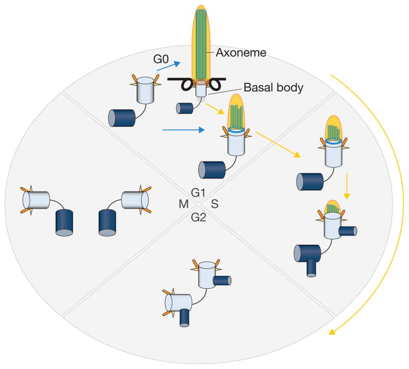
The primary cilium is an antenna-like, immotile organelle present on most types of mammalian cells, which interprets extracellular signals that regulate growth and development. Although once considered a vestigial organelle, the primary cilium is now the focus of considerable interest. We now know that ciliary defects lead to a panoply of human diseases, termed ciliopathies, and the loss of this organelle may be an early signature event during oncogenic transformation. Ciliopathies include numerous seemingly unrelated developmental syndromes, with involvement of the retina, kidney, liver, pancreas, skeletal system and brain. Recent studies have begun to clarify the key mechanisms that link cilium assembly and disassembly to the cell cycle, and suggest new possibilities for therapeutic intervention.


Conflict of interest statement

The authors declare no competing financial interests.

Figures

Figure 1
Figure 1. Linkage of the centrosome–cilium cycle to the cell cycle
Primary cilia assemble specifically when cells exit the cell cycle and become quiescent or differentiate. Cells are also competent to form cilia in G1. Phases of the cell cycle are indicated, and blue and yellow arrows indicate cilium assembly and disassembly, respectively. Only the mother centriole (light blue) can initiate ciliogenesis. The daughter centrioles are shown in dark blue. During the process of ciliogenesis, an axoneme is assembled. This microtubular structure (indicated with parallel green rods) is disassembled as cells progress towards S phase, concomitant with remodelling of the distal end of the basal body (aqua ring). During S phase, centrosomes commence duplication, at which point cilia have largely disassembled. After mitosis, centrosomes are again competent to assemble primary cilia, either in G0 or in early G1 phase.
Figure 2
Figure 2. Key players and events in cilium assembly
Cilium assembly proceeds through a series of orchestrated and well-defined stages (labelled I–IV), resulting in the dramatic remodelling of the maternal centriole. Exit from the cell cycle initiates the recruitment of Rabin8, a guanine nucleotide exchange factor (GEF), to the pericentriolar recycling endosome, whereupon it is activated by Rab11, setting off a cascade that ultimately activates Rab8a vesicles for recruitment and docking to the distal appendages (DA, orange antennae) of mother centrioles (I–II). Shortly after quiescence is induced, small vesicles (distal appendage vesicles, yellow) associated with Rab11 assemble around the DA, docking at the appendages (II). Distal appendage vesicles fuse to form ciliary vesicles through the action of the Ehd1 protein (II–III). After ciliary vesicle (CV) formation, the distal ends of mother centrioles/basal bodies are remodelled (concentric rings) through the action of: the kinase TTBK2, the balance of PtdIns (modulated by INPP5E and PIPKIγ), and CP110 removal, which releases the inhibition of microtubule growth and axonemal extension (III–IV). A second kinase, MARK4, could collaborate with TTBK2 to remove CP110 from distal ends. Vesicular Rab11 probably activates Rab8 for subsequent elongation of the ciliary membrane. After removal of CP110, the transition zone (TZ), which functions as a ciliary gate, is assembled, and IFT helps transport proteins through this gate.
Figure 3
Figure 3. Cilium disassembly
Serum growth factors trigger cilium disassembly in G1 phase through the concerted actions of two kinesins (Kif2a and Kif24) and destabilization of acetylated tubulins in the ciliary axoneme. The initiation of ciliary disassembly in early G1 requires the inhibition of axoneme extension (Plk1/Kif2a) and recruitment and activation of AurA/HEF1, which activates HDAC6 (H, green circles) and tubulin de-acetylation; this, in turn, leads to disassembly (steps I–III). Pitchfork (Pifo) and trichoplein collaborate to activate AurA, and Ndel1 stabilizes trichoplein at basal bodies to suppress ciliogenesis. Plk1-mediated phosphorylation of Kif2a stimulates its ability to depolymerize axonemal microtubules shortly after mitogen stimulation. At a later point, Nek2 is expressed and associates with Kif24, phosphorylating and activating this second de-polymerizing kinesin. Activation of Kif24 provides a sustained block to re-ciliation throughout S/G2/M (step IV), thereby ensuring that centrioles remain competent to replicate in S phase and assemble a mitotic spindle in M phase.

References

    1. Kobayashi T, Dynlacht BD. Regulating the transition from centriole to basal body. J Cell Biol. 2011;193:435–444. - PMC - PubMed
    1. Nigg EA, Stearns T. The centrosome cycle: centriole biogenesis, duplication and inherent asymmetries. Nat Cell Biol. 2011;13:1154–1160. - PMC - PubMed
    1. Kozminski KG, Johnson KA, Forscher P, Rosenbaum JL. A motility in the eukaryotic flagellum unrelated to flagellar beating. Proc Natl Acad Sci USA. 1993;90:5519–5523. - PMC - PubMed
    1. Pazour GJ, et al. Chlamydomonas IFT88 and its mouse homologue, polycystic kidney disease gene tg737, are required for assembly of cilia and flagella. J Cell Biol. 2000;151:709–718. - PMC - PubMed
    1. Goto H, Inoko A, Inagaki M. Cell cycle progression by the repression of primary cilia formation in proliferating cells. Cell Mol Life Sci. 2013;70:3893–3905. - PMC - PubMed

Publication types

LinkOut - more resources