. 2016 Aug 9;7(32):51784-51814.
doi: 10.18632/oncotarget.10108.

Long non-coding RNA NEAT1 promotes non-small cell lung cancer progression through regulation of miR-377-3p-E2F3 pathway

Affiliations

Long non-coding RNA NEAT1 promotes non-small cell lung cancer progression through regulation of miR-377-3p-E2F3 pathway

Chengcao Sun et al. Oncotarget. .

Abstract

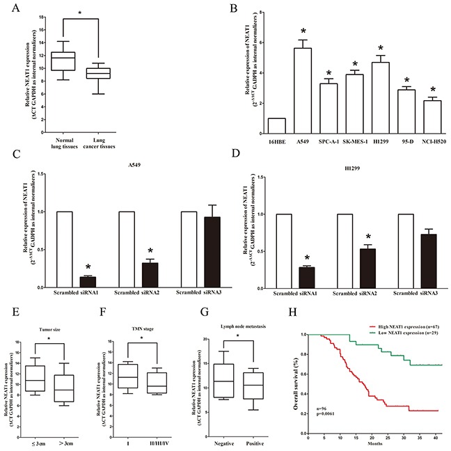
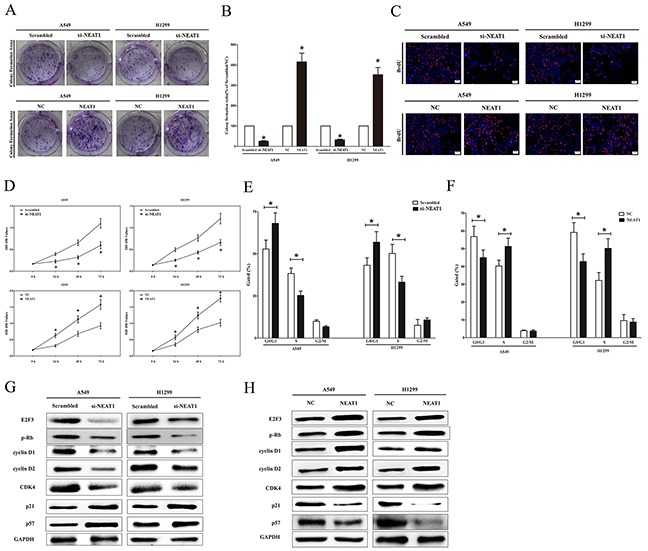
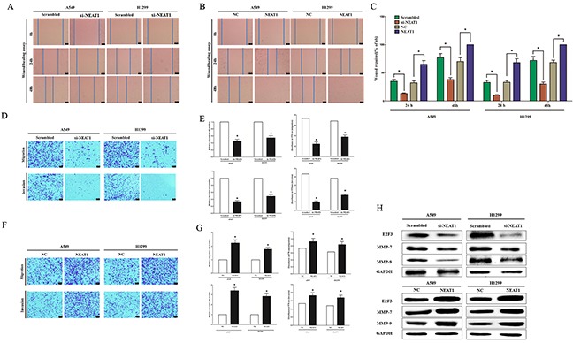
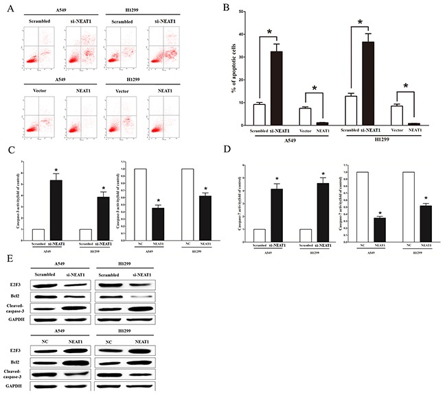
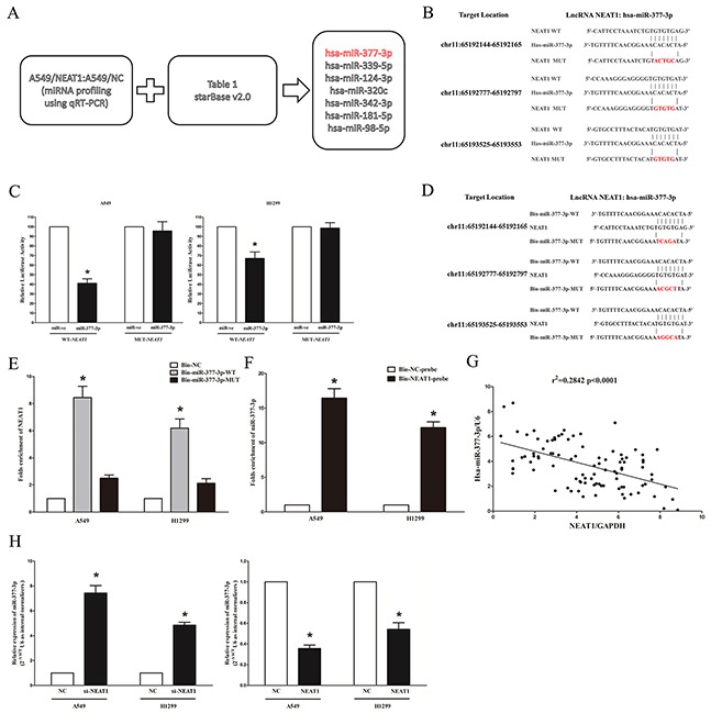
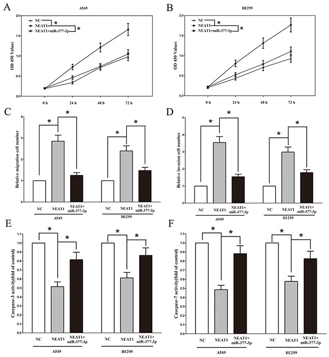
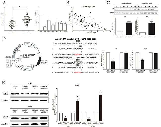
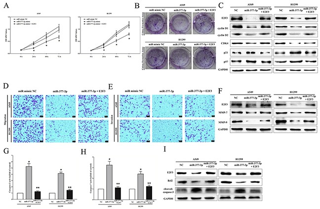
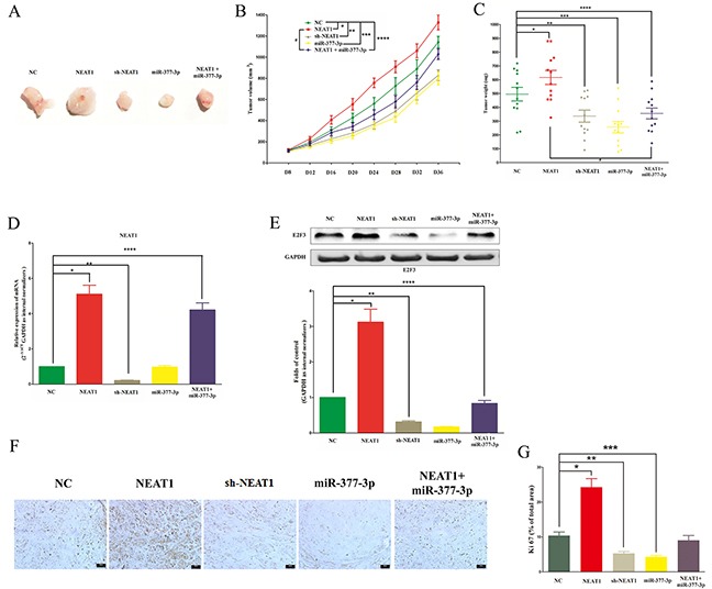
Recently, the long non-coding RNA (lncRNA) NEAT1 has been identified as an oncogenic gene in multiple cancer types and elevated expression of NEAT1 was tightly linked to tumorigenesis and cancer progression. However, the molecular basis for this observation has not been characterized in progression of non-small cell lung cancer (NSCLC). In our studies, we identified NEAT1 was highly expressed in patients with NSCLC and was a novel regulator of NSCLC progression. Patients whose tumors had high NEAT1 expression had a shorter overall survival than patients whose tumors had low NEAT1 expression. Further, NEAT1 significantly accelerates NSCLC cell growth and metastasis in vitro and tumor growth in vivo. Additionally, by using bioinformatics study and RNA pull down combined with luciferase reporter assays, we demonstrated that NEAT1 functioned as a competing endogenous RNA (ceRNA) for hsa-miR-377-3p, antagonized its functions and led to the de-repression of its endogenous targets E2F3, which was a core oncogene in promoting NSCLC progression. Taken together, these observations imply that the NEAT1 modulated the expression of E2F3 gene by acting as a ceRNA, which may build up the missing link between the regulatory miRNA network and NSCLC progression.

Keywords: E2F3; hsa-miRNA-377-3p (miR-377-3p); long non-coding RNA NEAT1 (lncRNA NEAT1); non-small cell lung cancer (NSCLC); tumorigenesis.


Conflict of interest statement

The authors declare no conflict of interest.

Figures

Figure 1
Figure 1. Relative NEAT1 expression in non-small cell lung cancer tissues and cell lines, and its clinical significance
A. Relative expression of NEAT1 expression in NSCLC tissues (n = 96) and in paired adjacent normal tissues (n = 96). NEAT1 expression was examined by qPCR and normalized to GAPDH expression. (shown as ΔCT). B. Relative expression of NEAT1 expression in NSCLC cell lines and normal HELF lung epidermal cell. C-D. Relative NEAT1 expression in A549 and H1299 cells after transfecting with si-NEAT1, namely, siRNA1, siRNA2 and siRNA3. NEAT1 expression was examined by qPCR and normalized to GAPDH expression (shown as 2−ΔΔCT). E-G. NEAT1 expression was significantly higher in patients with big tumor size, advanced clinical stage and lymph nodes metastasis. NEAT1 expression was examined by qPCR and normalized to GAPDH expression. (shown as ΔCT). H. The Kaplan-Meier survival analysis indicated that NEAT1 high expression (red line, n=67) has a worse overall survival compared to the low expression subgroup (green line, n=29). *P < 0.05. Means ± SEM are shown. Statistical analysis was conducted using student t-test.
Figure 2
Figure 2. NEAT1 promotes tumor NSCLC cell growth in vitro
A. Shown are representative photomicrographs of colony formation assay after transfection for fourteen days. B. Statistical analysis of colony formation assay. C. Shown are representative photomicrographs of BrdU staining in A549 and H1299 cells after transfection. Bar = 100 μm. D. CCK8 assays of A549 and H1299 cells after transfection. F. Cell-cycle analysis was performed after transfection for forty eight hours. The DNA content was quantified by flow cytometric analysis. G. Expression of E2F3, p-Rb, cyclin D1, cyclin D2, CDK4, p21 and p57 protein in transfected A549 and H1299 cells. Assays were performed in triplicate. *P < 0.05. Means ± SEM are shown. Statistical analysis was conducted using student t-test.
Figure 3
Figure 3. NEAT1 promotes NSCLC cell migration and invasion in vitro
A-B. Shown are representative photomicrographs of “wound healing assay” in A549 and H1299 cells after transfection for 0 hour, twenty hours and forty eight hours. Bar = 50 μm. C. Statistical analysis of “wound healing assay”. D-G. A549 and H1299 cells were loaded onto the top well of a transwell inserts for cell migration or invasion assay. After twenty four hours, cells that migrated to the bottom chamber containing serum-supplemented medium were stained with 0.1% crystal violet, visualized under a phase-contrast microscope, and photographed. Bar = 50 μm. Total number of cells in five fields was counted manually. H. Expression of E2F3, MMP-7 and MMP-9 protein in A549 and H1299 cells after transfection. Assays were performed in triplicate. *P < 0.05. Means ± SEM are shown. Statistical analysis was conducted using student t-test.
Figure 4
Figure 4. NEAT1 inhibits NSCLC cell apoptosis in vitro
A. Shown are representative photomicrographs of flow cytometric analysis. B. Statistical analysis of flow cytometric analysis. C-D. Quantitative representation of caspase-3 and caspase-7 activity in A549 and H1299 cells after transfection for forty eight hours. E. Western-blot of E2F3, Bcl2 and cleaved-caspase-3 protein in A549 and H1299 cells after transfection. Assays were performed in triplicate. *P < 0.05. Means ± SEM are shown. Statistical analysis was conducted using student t-test.
Figure 5
Figure 5. NEAT1 is a direct target of miR-377-3p
A. Screen of the candidate miRNAs that interacted with NEAT1 by real-time PCR-based miRNA expression profiling and starBase (v2.0). Co-analysis of the down-regulated miRNAs in stable overexpression NEAT1/A549 cells compared to the control A549 cells and the miRNA list that potentially target NEAT1 predicted by starBase (v2.0), shown in Table 1, we got seven candidates. B. Sequence alignment of miR-377-3p with the putative binding sites within the wild-type regions of NEAT1. C. The luciferase report assay demonstrated that overexpression of miR-377-3p could reduce the intensity of fluorescence in A549 and H1299 cells transfected with the NEAT1-WT vector, while had no effect on the NEAT1-MUT vector. D.WT and the mutated forms of miR-377-3p sequence are shown. E. Detection of NEAT1 using qRT-PCR in the sample pulled down by biotinylated miR-377-3p. F. Detection of miR-377-3p using qRT-PCR in the sample pulled down by biotinylated NEAT1 probe. G. The correlation between NEAT1 mRNA and miR-377-3p expression in 96 lung cancer tissues. H. Detection of miR-377-3p using qRT-PCR in the si-NEAT1 or NEAT1 overexpression A549 and H1299 cell lines compared with control group. Assays were performed in triplicate. *P < 0.05. Means ± SEM are shown. Statistical analysis was conducted using student t-test.
Figure 6
Figure 6. NEAT1's oncogenic activity is in part through negative regulation of miRNA-377-3p in NSCLC cells
Up-regulated miR-377-3p in A549 and H1299 cells, which stably overexpressed NEAT1, largely reversed the favorable effects of NEAT1 on cell proliferation A-B. migration C. and invasion D. Moreover, overexpression of miR-377-3p largely increased the cell apoptosis inhibited by NEAT1 E-F. Assays were performed in triplicate. *P < 0.05. Means ± SEM are shown. Statistical analysis was conducted using student One-Way ANOVA test.
Figure 7
Figure 7. E2F3 proto-oncogene is a target of miR-377-3p at specific 3′-UTR sites
A. miR-377-3p is lowly expressed in primary human lung cancer tissues (n=96) and NSCLC cell lines. B. Scatter plots showing the inverse association between miR-377-3p level and E2F3 mRNA expression in primary human lung cancer tissues (n=96). C. E2F3 is up-regulated in primary human lung cancer tissues (n=96). D. Left pmiR-RB-REPORT™ dual-luciferase reporter vector. Middle The 3′-UTR of E2F3 harbors two miR-377-3p cognate sites. Right Relative luciferase activity of reporter plasmids carrying wild-type or mutant E2F3 3′-UTR in A549 and H1299 cells co-transfected with negative control (NC) or miR-377-3p mimic. E. miR-377-3p suppressed the protein expression of E2F3. Assays were performed in triplicate. *P < 0.05. Means ± SEM are shown. Statistical analysis was conducted using student t-test.
Figure 8
Figure 8. miR-377-3p exerts tumor suppressor function through down-regulation of E2F3 in NSCLC cell lines
A-B. CCK8 and colony formation assays demonstrated E2F3 reversed the growth inhibitory role of miR-377-3p in A549 and H1299 cells. C. Protein expression of E2F3, cyclin D1, cyclin D2, CDK4, p21, and p57 after transfection. D-E. Transwell migration/invasion assays demonstrated that E2F3 reversed the inhibitory role of miR-377-3p on migration and invasion in A549 and H1299 cells. F. Protein expression of E2F3, MMP-7, and MMP-9 after transfection. G-H. Caspase-3 and caspase-7 activity assays demonstrated that E2F3 reversed the favorable role of miR-377-3p on apoptosis in A549 and H1299 cells. I. Protein expression of E2F3, Bcl2, and cleaved-caspase-3 after transfection. Assays were performed in triplicate. *P < 0.05, **P < 0.05. Means ± SEM are shown. Statistical analysis was conducted using student One-Way ANOVA test.
Figure 9
Figure 9. NEAT1 promotes NSCLC cell growth in vivo by inhibiting miR-377-3p/E2F3 axis
A-C. Tumor size, volume, and weight of subcutaneous implantation models of A549 cell are shown. D. NEAT1 expression in tumors isolated from NC, NEAT1, sh-NEAT1, miR-377-3p, and NEAT1+miR-377-3p groups. E. The protein expression of E2F3 in tumors isolated from NC, NEAT1, sh-NEAT1, miR-377-3p, and NEAT1+miR-377-3p groups. F. Immunohistochemistry of Ki67 in tumors isolated from NC, NEAT1, sh-NEAT1, miR-377-3p, and NEAT1+miR-377-3p groups. G. Statistics of Ki 67 IHC. Assays were performed in triplicate. *P < 0.05, **P < 0.05, ***P < 0.05, ****P < 0.05. Means ± SEM are shown. Statistical analysis was conducted using student One-Way ANOVA test.

References

    1. IARC Globocan 2012 Cancer Fact Sheet: Lung Cancer Mortality Worldwide in 2012.
    1. Sun C, Li S, Yang C, Xi Y, Wang L, Zhang F, Li D. MicroRNA-187-3p mitigates non-small cell lung cancer (NSCLC) development through down-regulation of BCL6. Biochem Biophys Res Commun. 2016;471:82–88. - PubMed
    1. Laskin JJ, Sandler AB. State of the art in therapy for non-small cell lung cancer. Cancer Invest. 2005;23:427–442. - PubMed
    1. Tong AW. Small RNAs and non-small cell lung cancer. Curr Mol Med. 2006;6:339–349. - PubMed
    1. Sekido Y, Fong KM, Minna JD. Molecular genetics of lung cancer. Annu Rev Med. 2003;54:73–87. - PubMed

MeSH terms