Case Reports
. 2016 Aug;80(2):294-300.
doi: 10.1002/ana.24715. Epub 2016 Jul 15.

CTLA4 as Immunological Checkpoint in the Development of Multiple Sclerosis

Affiliations
Case Reports

CTLA4 as Immunological Checkpoint in the Development of Multiple Sclerosis

Lisa Ann Gerdes et al. Ann Neurol. 2016 Aug.

Abstract

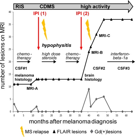
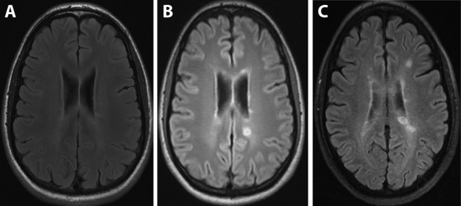
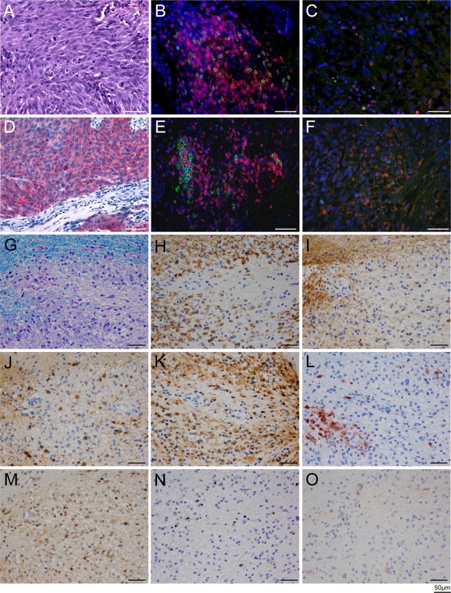
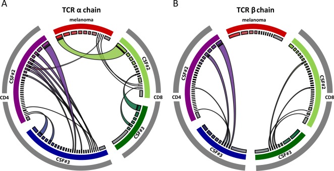
We investigated a patient who developed multiple sclerosis (MS) during treatment with the CTLA4-blocking antibody ipilimumab for metastatic melanoma. Initially he showed subclinical magnetic resonance imaging (MRI) changes (radiologically isolated syndrome). Two courses of ipilimumab were each followed by a clinical episode of MS, 1 of which was accompanied by a massive increase of MRI activity. Brain biopsy confirmed active, T-cell type MS. Quantitative next generation sequencing of T-cell receptor genes revealed distinct oligoclonal CD4(+) and CD8(+) T-cell repertoires in the primary melanoma and cerebrospinal fluid. Our results pinpoint the coinhibitory molecule CTLA4 as an immunological checkpoint and therapeutic target in MS. Ann Neurol 2016;80:294-300.


Figures

Figure 1
Figure 1
Synopsis of the clinical course. An overview of the course, treatments, serial magnetic resonance imaging (MRI) changes, and time points of cerebrospinal fluid (CSF) samplings is presented. Before ipilimumab (IPI) treatment was begun, the patient had subclinical MRI activity fulfilling the criterion of dissemination in time (radiologically isolated syndrome (RIS)). Each course of ipilimumab was followed by a clinical episode of multiple sclerosis (MS), the first of which marked transition to clinically definite MS (CDMS). At the time of melanoma diagnosis and staging (RIS), cranial MRI showed a total of 11 fluid‐attenuated inversion recovery (FLAIR) and 3 Gadolinium enhancing (Gd+) lesions; 4 months after the second course of ipilimumab, MRI showed 24 cumulative FLAIR and 9 new Gd+ lesions; 10 months after the second course of ipilimumab, MRI showed 37 cumulative FLAIR and 6 Gd+ lesions.
Figure 2
Figure 2
Course of magnetic resonance imaging (MRI). Representative serial cranial MRI scans (fluid‐attenuated inversion recovery) were performed at the stage of radiologically isolated syndrome before ipilimumab treatment (A), and 4 months (B) or 10 months (C) after the second course of ipilimumab. The time points of MRI A to C are indicated in Figure 1.
Figure 3
Figure 3
Histology of the primary melanoma (A–F) and active demyelinating multiple sclerosis (MS) lesion (G–O). (A–F) Hematoxylin and eosin staining of the primary melanoma is shown in A. Melanoma cells exhibit strong cytoplasmic staining for Melan‐A (D). Double‐stainings for CD3 (red) and CD8 (green; B, C), and double staining for CD3+ (red) and CD20 (green; E, F) show infiltration of immune cells into the epidermis (B, E), as well as scattered T and B cells in the tumor (C, F). The ratios of total (CD3+) T cells to cytotoxic (CD8+) T cells, and total (CD3+) T cells to CD20+ B cells, were 3.4:1 and 3:1, respectively. Nuclei are stained in blue with DAPI. (G–O) Brain biopsy specimen of active demyelinating MS lesion. Region of active demyelination with numerous foamy macrophages containing myelin debris (G, stained with Luxol fast blue/periodic acid–Schiff; H, stained for myelin proteolipid protein) is shown. The foamy macrophages also contained fragments of myelin oligodendrocyte glycoprotein (I). J shows Nogo A–positive, viable oligodendrocytes in the demyelinated area; K shows dense infiltration of foamy macrophages and activated microglia with KiM1P immunohistochemistry. Some foamy macrophages are positive for the early macrophage activation marker MRP14 (L). Scattered CD3+ T cells are present in the demyelinated area (M); most infiltrating T cells are CD8+ (N). The foamy macrophages stain negative for complement C9neo, as is typical for pattern 1 MS lesions10 (O). All scale bars represent 50 μm.
Figure 4
Figure 4
Next generation sequencing of the T‐cell receptor (TCR) repertoire in the primary melanoma and 2 consecutive cerebrospinal fluid (CSF) samples (CSF#2 and CSF#3; cf Fig 1). (A) The TCR α‐chain repertoire is shown on the left. (B) The TCR ß‐chain repertoire is shown on the right. The TCR repertoire of the melanoma is highlighted in red at the top of each circle; the CD4+ TCR repertoire of CSF#2 and CSF#3 is shown in purple and blue on the left of each circle, and the CD8+ TCR repertoire of CSF#2 and CSF#3 is shown in light and dark green on the right of each circle. The widths of the colored segments of the inner circles indicate the relative abundance of each T‐cell clone; the polyclonal background is depicted in gray (inner circle). Expanded T‐cell clones shared between the compartments are visualized as semicircular connections. There are several overlaps, mostly of CD8+ TCR α‐chains, between the melanoma and CSF#2, but not CSF#3 (A). Furthermore, there are overlaps between CSF#2 and CSF#3 at all levels (TCR α‐ and ß‐chains; CD4+ and CD8+ T cells; A and B). The CSF‐specific, persistent clonal expansions likely contribute to multiple sclerosis in this patient. Images were generated using CIRCOS software (http://circos.ca/).

References

    1. Gourraud PA, Harbo HF, Hauser SL, Baranzini SE. The genetics of multiple sclerosis: an up‐to‐date review. Immunol Rev 2012;248:87–103. - PMC - PubMed
    1. Dendrou CA, Fugger L, Friese MA. Immunopathology of multiple sclerosis. Nat Rev Immunol 2015;15:545–558. - PubMed
    1. Nylander A, Hafler DA. Multiple sclerosis. J Clin Invest 2012;122:1180–1188. - PMC - PubMed
    1. Sawcer S, Franklin RJ, Ban M. Multiple sclerosis genetics. Lancet Neurol 2014;13:700–709. - PubMed
    1. Sawcer S, Hellenthal G, Pirinen M, et al. Genetic risk and a primary role for cell‐mediated immune mechanisms in multiple sclerosis. Nature 2011;476:214–219. - PMC - PubMed

Publication types

MeSH terms