. 2016 Oct;33(10):2540-51.
doi: 10.1007/s11095-016-1978-1. Epub 2016 Jun 28.

Targeting of Micelles and Liposomes Loaded with the Pro-Apoptotic Drug, NCL-240, into NCI/ADR-RES Cells in a 3D Spheroid Model

Affiliations

Targeting of Micelles and Liposomes Loaded with the Pro-Apoptotic Drug, NCL-240, into NCI/ADR-RES Cells in a 3D Spheroid Model

Bhushan S Pattni et al. Pharm Res. 2016 Oct.

Erratum in

Abstract

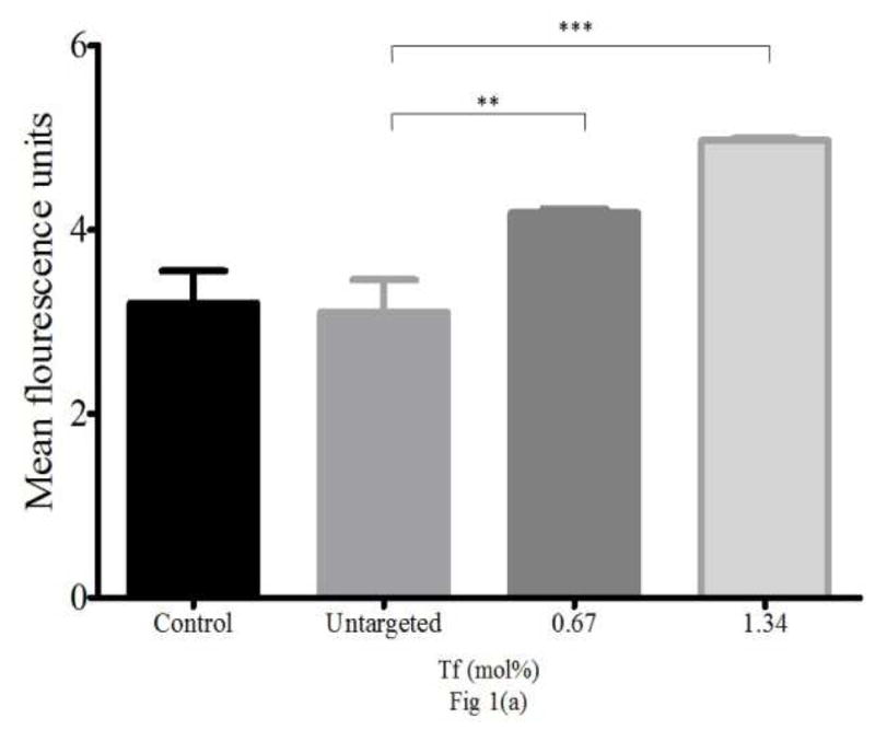
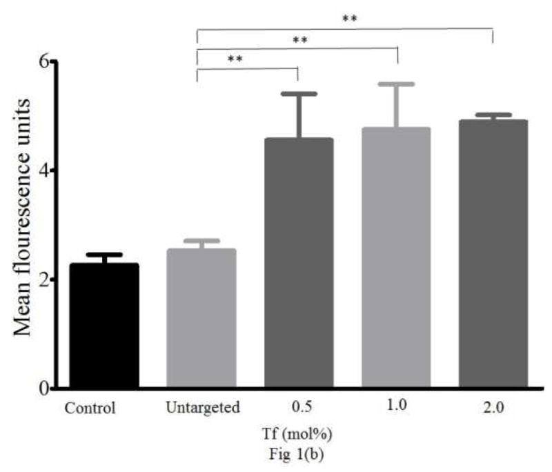
Purpose: To develop transferrin (Tf)-targeted delivery systems for the pro-apoptotic drug, NCL-240, and to evaluate the efficacy of this delivery system in ovarian cancer NCI/ADR-RES cells, grown in vitro in a 3D spheroid model.

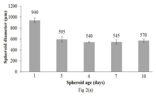
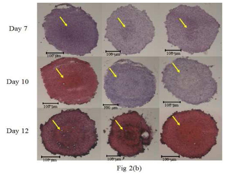
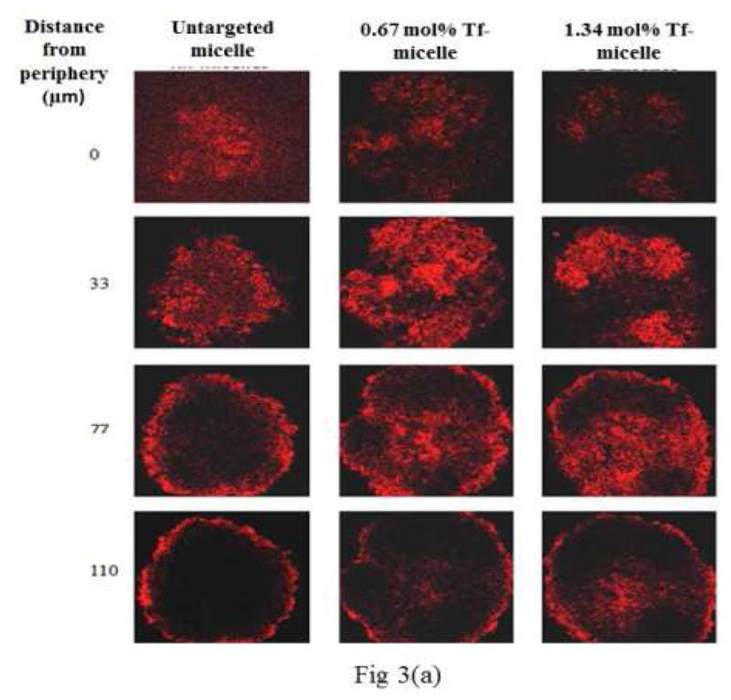
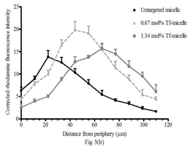
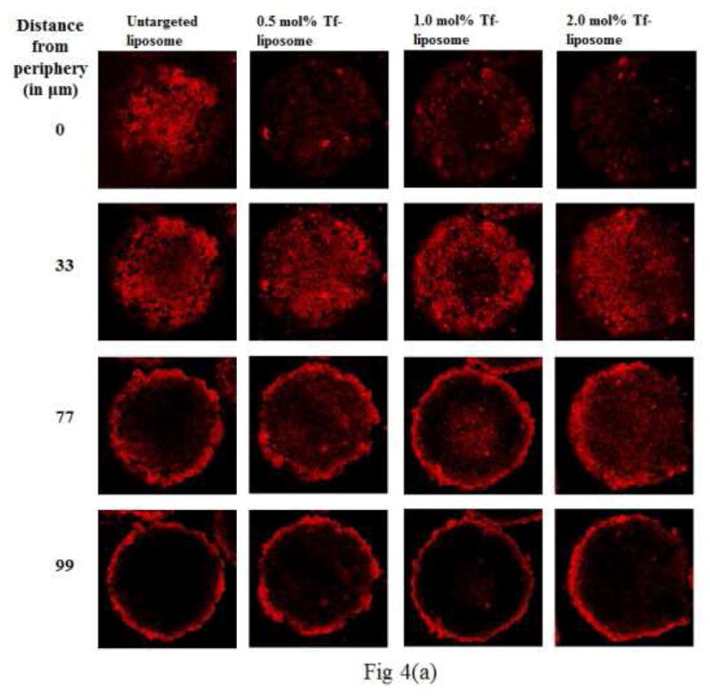
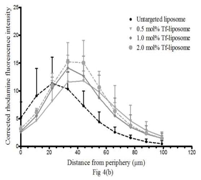
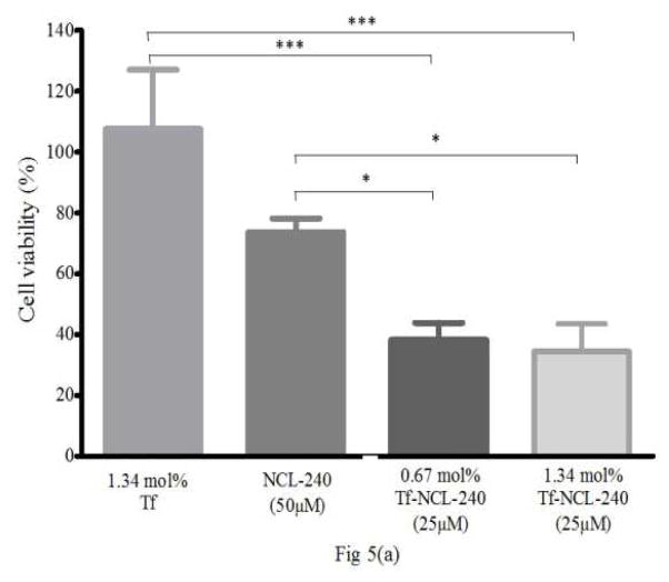
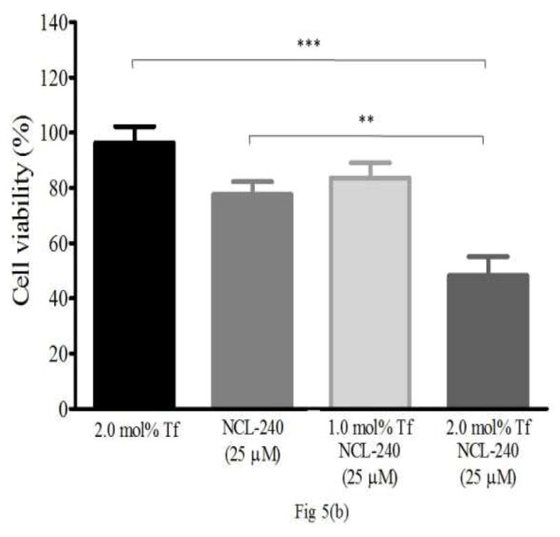
Methods: Tf-targeted PEG-PE-based micellar and ePC/CHOL-based liposomal delivery systems for NCL-240 were prepared. NCI/ADR-RES cells were used to generate spheroids by a non-adhesive liquid overlay technique. Spheroid growth and development were monitored by size (diameter) analysis and H&E staining. The targeted formulations were compared to untargeted ones in terms of their degree of spheroid association and penetration. A cell viability analysis with NCL-240-loaded micelles and liposomes was performed to assess the effectiveness of Tf-targeting.

Results: Tf-targeted polymeric micelles and Tf-targeted liposomes loaded with NCL-240 were prepared. NCI/ADR-RES cells generated spheroids that demonstrated the presence of a distinct necrotic core along with proliferating cells in the spheroid periphery, partly mimicking in vivo tumors. The Tf-targeted micelles and liposomes had a deeper spheroid penetration as compared to the untargeted delivery systems. Cell viability studies using the spheroid model demonstrated that Tf-mediated targeting markedly improved the cytotoxicity profile of NCL-240.

Conclusion: Transferrin targeting enhanced delivery and effectiveness of micelles and liposomes loaded with NCL-240 against NCI/ADR-RES cancer cells in a 3D spheroid model.

Keywords: NCL-240; cancer spheroids; liposomes; micelles; transferrin-targeted drug delivery.


Figures

Figure 1
Figure 1. Cellular association of Tf-targeted (a) micelles and (b) liposomes measured using FACS
(a) Association of Tf-conjugated micelles with NCI/ADR-RES. (b) Association of Tf-targeted liposomes with NCI/ADR-RES cells (n=3, Mean ± S.D., ** p≤0.01, *** p≤0.001).
Figure 1
Figure 1. Cellular association of Tf-targeted (a) micelles and (b) liposomes measured using FACS
(a) Association of Tf-conjugated micelles with NCI/ADR-RES. (b) Association of Tf-targeted liposomes with NCI/ADR-RES cells (n=3, Mean ± S.D., ** p≤0.01, *** p≤0.001).
Figure 2
Figure 2. NCI/ADR-RES spheroid growth
(a) Spheroid diameter with age (Mean ± S.D., n=3) (b) H&E stained sections of NCI/ADR-RES spheroids by fluorescence microscopy. From day 7 onward, the formation of a distinctly stained core (indicated by arrow) became visible. Day 10 spheroids showed 2 distinct layers consisting of a proliferative outer shell and the necrotic inner core. By day 12, the spheroids had begun shedding cells on the periphery.
Figure 3
Figure 3. Micelle penetration of spheroids
(a) Z-stack images taken by confocal microscopy showing penetration of untargeted and Tf-targeted rhodamine (Rh)-labeled micelles in 4 days old NCI/ADR-RES spheroids. The images are of spheroid sections at 0, 33, 77 and 110 μm distance from the periphery. The red color indicates the presence of Rh-labeled micelles. The micelle association with the deeper layers of the spheroid was higher when targeted. (b) The corrected rhodamine fluorescence intensity represents the micelle association of targeted and untargeted micelles in NCI/ADR-RES spheroids from the periphery to the inner layers.
Figure 4
Figure 4. Liposome penetration in spheroids
(a) Z-stack images taken by confocal microscopy show penetration of untargeted and Tf-targeted rhodamine (Rh)-labeled liposomes in 4 days old NCI/ADR-RES spheroids. The images are of spheroid sections at 0, 33, 77 and 99 μm distance from the periphery. The red color indicates the presence of Rh-labeled liposomes. The association with the deeper layers of the spheroid was higher with Tf-targeted liposomes. (b) The corrected rhodamine fluorescence intensity vs distance from the periphery quantitates the liposome association of targeted and untargeted liposomes in NCI/ADR-RES spheroids.
Figure 5
Figure 5. Cell viability in spheroids after treatment with NCL-240-loaded (a) micelles and (b) liposomes
Cell viability in spheroids after 4 h treatment with NCL-240-micelles (a). With a 25 μM dose of NCL-240 in Tf-targeted (0.67 or 1.34 mol%) micelles, significant cytotoxicity occurred compared to a 50 μM dose of untargeted micellar NCL-240 (a). Cell viability in spheroids after 2 h treatment with NCL-240-liposomes (b). A 25 μM dose of NCL-240 in 2.0 mol% Tf-targeted in liposomes significantly increased cytotoxicity compared to untargeted NCL-240 liposomes. (n=3, Mean ± S.D., * p≤0.05, ** p≤0.01, *** p≤0.001)

References

    1. Fresno Vara JA, Casado E, de Castro J, Cejas P, Belda-Iniesta C, Gonzalez-Baron M. PI3K/Akt signalling pathway and cancer. Cancer treatment reviews. 2004 Apr;30(2):193–204. - PubMed
    1. Courtney KD, Corcoran RB, Engelman JA. The PI3K pathway as drug target in human cancer. Journal of clinical oncology : official journal of the American Society of Clinical Oncology. 2010 Feb 20;28(6):1075–83. - PMC - PubMed
    1. Liu P, Cheng H, Roberts TM, Zhao JJ. Targeting the phosphoinositide 3-kinase pathway in cancer. Nature reviews Drug discovery. 2009 Aug;8(8):627–44. - PMC - PubMed
    1. Miao B, Skidan I, Yang J, Lugovskoy A, Reibarkh M, Long K, et al. Small molecule inhibition of phosphatidylinositol-3,4,5-triphosphate (PIP3) binding to pleckstrin homology domains. Proceedings of the National Academy of Sciences of the United States of America. 2010 Nov 16;107(46):20126–31. - PMC - PubMed
    1. Riehle RD, Cornea S, Degterev A, Torchilin V. Micellar formulations of pro-apoptotic DM-PIT-1 analogs and TRAIL in vitro and in vivo. Drug delivery. 2013 Feb;20(2):78–85. - PubMed

Publication types

MeSH terms