. 2017 Jan;28(1):218-229.
doi: 10.1681/ASN.2015121376. Epub 2016 Jun 27.

Ferroptosis, but Not Necroptosis, Is Important in Nephrotoxic Folic Acid-Induced AKI

Affiliations

Ferroptosis, but Not Necroptosis, Is Important in Nephrotoxic Folic Acid-Induced AKI

Diego Martin-Sanchez et al. J Am Soc Nephrol. 2017 Jan.

Abstract

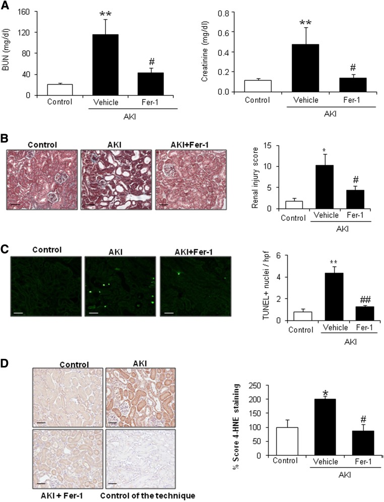
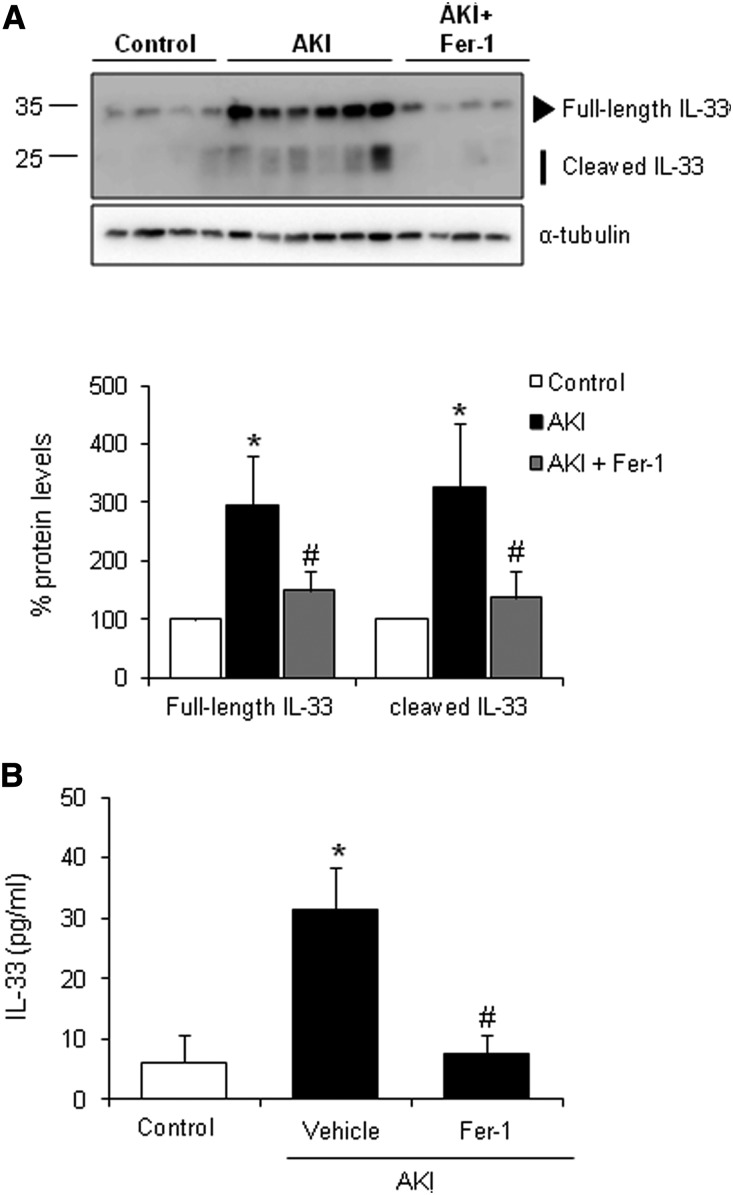
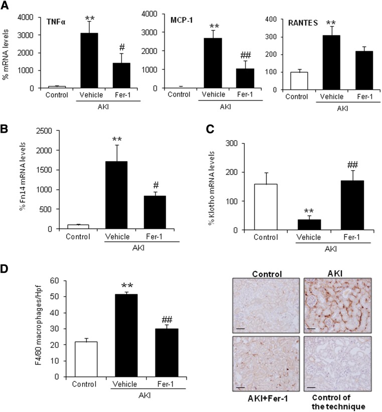
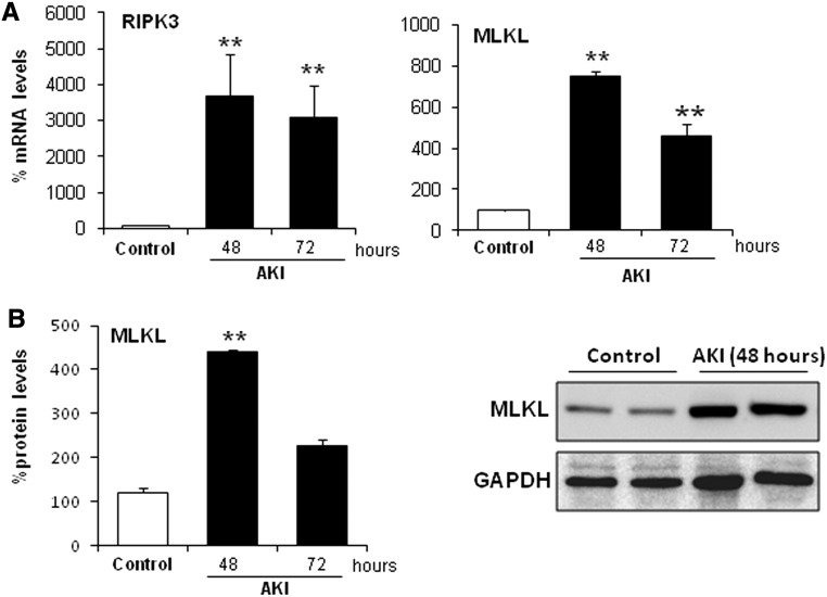
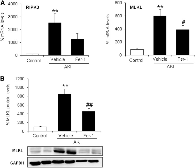
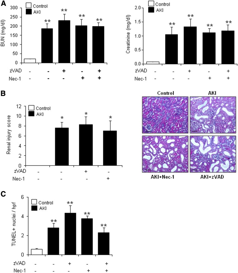
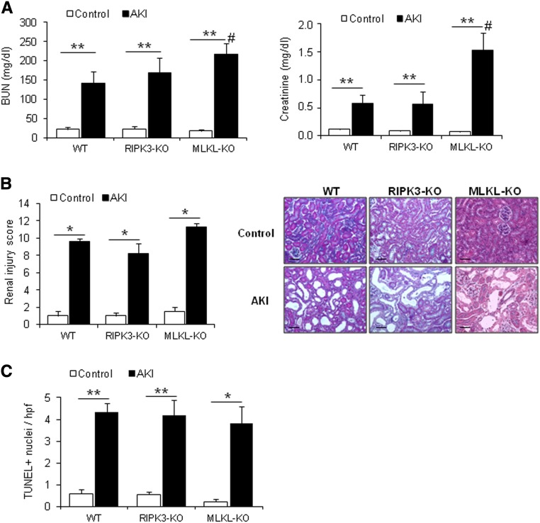
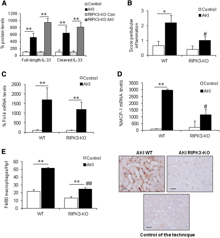
AKI is histologically characterized by necrotic cell death and inflammation. Diverse pathways of regulated necrosis have been reported to contribute to AKI, but the molecular regulators involved remain unclear. We explored the relative contributions of ferroptosis and necroptosis to folic acid (FA)-induced AKI in mice. FA-AKI in mice associates with lipid peroxidation and downregulation of glutathione metabolism proteins, features that are typical of ferroptotic cell death. We show that ferrostatin-1 (Fer-1), an inhibitor of ferroptosis, preserved renal function and decreased histologic injury, oxidative stress, and tubular cell death in this model. With respect to the immunogenicity of ferroptosis, Fer-1 prevented the upregulation of IL-33, an alarmin linked to necroptosis, and other chemokines and cytokines and prevented macrophage infiltration and Klotho downregulation. In contrast, the pancaspase inhibitor zVAD-fmk did not protect against FA-AKI. Additionally, although FA-AKI resulted in increased protein expression of the necroptosis mediators receptor-interacting protein kinase 3 (RIPK3) and mixed lineage domain-like protein (MLKL), targeting necroptosis with the RIPK1 inhibitor necrostatin-1 or genetic deficiency of RIPK3 or MLKL did not preserve renal function. Indeed, compared with wild-type mice, MLKL knockout mice displayed more severe AKI. However, RIPK3 knockout mice with AKI had less inflammation than their wild-type counterparts, and this effect associated with higher IL-10 concentration and regulatory T cell-to-leukocyte ratio in RIPK3 knockout mice. These data suggest that ferroptosis is the primary cause of FA-AKI and that immunogenicity secondary to ferroptosis may further worsen the damage, although necroptosis-related proteins may have additional roles in AKI.

Keywords: IL-33; acute renal failure; cell death; ferroptosis; inflammation; renal proximal tubule cell.


Figures

Figure 1.
Figure 1.
Fer-1 prevented renal injury in FA-induced AKI. C57/BL6 mice were pretreated with Fer-1 for 30 minutes followed by AKI induction by an FA overdose and euthanasia at 48 hours. (A) Renal function was assessed by plasma creatinine and BUN levels. Fer-1 preserved renal function. Mean±SEM of seven mice per group. **P<0.01 versus control; #P<0.04 versus AKI alone. (B) Fer-1 decreased histologic injury in kidneys from mice exposed to FA. Representative image of Masson staining and quantification. Mean±SEM of seven mice per group. Original magnification, ×200. Scale bars, 50 μm. *P<0.02 versus control; #P<0.04 versus AKI alone. (C) Cell death was assessed by TUNEL staining (green). Fer-1 reduced TUNEL+ cells in FA–induced AKI kidneys. Representative image and quantification as mean±SEM of seven mice per group. Original magnification, ×400. Scale bars, 50 μm. **P<0.002 versus control; ##P<0.001 versus AKI alone. (D) 4-Hydroxynonenal (4-HNE) staining in mice with AKI. Fer-1 decreased 4-HNE levels. Mean±SEM of seven animals per group. Original magnification, ×200. Scale bars, 50 μm. *P<0.03 versus control; #P<0.02 versus AKI alone.
Figure 2.
Figure 2.
Fer-1 decreases IL-33 levels and activation in FA-induced AKI. IL-33 protein levels were measured in mice with AKI pretreated or not pretreated with Fer-1. (A) Kidney IL-33 upregulation and processing observed in AKI at 48 hours was prevented with Fer-1 treatment. Representative Western blot and quantification of IL-33 levels. Mean±SEM of seven animals per group. *P<0.01 versus control; #P<0.04 versus AKI. (B) Plasma IL-33 levels assayed by ELISA were incremented in AKI at 48 hours, and this was prevented by Fer-1. Mean±SEM of seven animals per group. *P<0.05 versus control; #P<0.03 versus AKI.
Figure 3.
Figure 3.
Renal inflammation associated with AKI is modulated by Fer-1. (A) Fer-1 prevented the increase in kidney TNFα, MCP-1, and RANTES mRNA levels at 48 hours of AKI. Mean±SEM of seven animals per group. **P<0.002 versus control; #P<0.03 versus AKI alone; ##P<0.003 versus AKI alone. (B and C) Quantification of kidney Fn14 and Klotho mRNA levels. Fer-1 prevented (B) the increase in Fn14 and (C) the decrease in Klotho mRNA expression in AKI at 48 hours. Mean±SEM of seven animals per group. **P<0.003 versus control; #P<0.03 versus AKI alone; ##P<0.003 versus AKI alone. (D) Fer-1 prevented the increase in F4/80–positive interstitial macrophages in AKI kidneys. Mean±SEM of seven animals per group. Original magnification, ×200. Scale bars, 50 μm. **P<0.002 versus control; ##P<0.001 versus AKI alone.
Figure 4.
Figure 4.
Necroptosis regulatory proteins are upregulated in FA-induced AKI. Kidney RIPK3 and MLKL levels were assessed in mice with AKI over time. (A) RIPK3 and MLKL mRNA expression is increased in a time-dependent fashion during AKI. Mean±SEM of five animals per group. **P<0.001 versus control. (B) Time course of MLKL protein expression in AKI assessed by Western blot. Quantification and representative Western blot. Mean±SEM of five animals per group. **P<0.004 versus control.
Figure 5.
Figure 5.
Fer-1 reduces levels of necroptosis regulatory proteins in AKI. (A) Fer-1 prevented the upregulation of kidney RIPK3 and MLKL mRNA expression in AKI. Mean±SEM of seven animals per group. **P<0.003 versus control; #P<0.03 versus AKI alone. (B) Representative Western blot and quantification of kidney MLKL protein. Mean±SEM of seven animals per group. **P<0.002 versus control; ##P<0.003 versus AKI.
Figure 6.
Figure 6.
Neither Nec-1 nor zVAD prevents renal injury in FA-induced AKI. C57/BL6 mice were pretreated with zVAD and/or Nec-1 for 30 minutes followed by AKI induction by an FA overdose and euthanasia at 48 hours. (A) Renal function was assessed by plasma creatinine and BUN levels in AKI. Neither zVAD nor Nec-1 preserved renal function. Mean±SEM of seven animals per group. **P<0.003 versus control. (B) Masson staining and quantification of histologic renal damage. Mean±SEM of seven animals per group. Original magnification, ×200. Scale bars, 50 μm. *P<0.04 versus control. (C) Quantification of TUNEL+ cells. Mean±SEM of seven animals per group. **P<0.004 versus control.
Figure 7.
Figure 7.
RIPK3 and MLKL are required for promoting regeneration after FA-induced AKI. AKI was induced in RIPK3-KO (n=8) and MLKL-KO (n=4–6) mice and their wild-type (WT) littermates (n=7). Mice were euthanized at 48 hours. (A) Renal function was assessed by plasma creatinine and BUN levels. Mean±SEM of four to eight animals per group. **P<0.003 versus control; #P<0.04 versus AKI WT. (B) Masson staining and quantification of histologic renal damage. Mean±SEM of four to eight animals per group. Original magnification, ×200. Scale bars, 50 μm. *P<0.02 versus control. (C) Quantification of TUNEL+ cells. Mean±SEM of four to eight animals per group. *P<0.02 versus control; **P<001 versus control.
Figure 8.
Figure 8.
Fer-1, but not zVAD, prevents renal injury in FA-induced AKI in RIPK3-KO mice. (A) RIPK3-KO mice were pretreated with Fer-1 (n=7) or vehicle (n=8) 30 minutes before inducing AKI by an FA overdose and euthanized 48 hours later. Renal function was assessed by plasma creatinine and BUN levels, and cell death was assessed by TUNEL staining. Quantification is expressed as mean±SEM of seven or eight mice per group. **P<0.002 versus control; #P<0.05 versus AKI alone. (B) M30 cytodeath staining to assess caspase activation in RIPK3-KO mice during FA-induced AKI. Representative image and quantification as mean±SEM of seven mice per group. WT, wild type. Original magnification, ×200. Scale bars, 50 μm. **P<0.002 versus WT mice. (C) RIPK3-KO mice were pretreated with zVAD (n=8) or vehicle (n=8) 30 minutes before AKI induction by an FA overdose and euthanized at 48 hours. Renal function was assessed by plasma creatinine and BUN levels, and cell death was assessed by TUNEL staining. Quantification as mean±SEM of eight mice per group. *P<0.02 versus control; **P<0.002 versus control.
Figure 9.
Figure 9.
RIPK3 deficiency prevented renal inflammation in FA-induced AKI. (A) Kidney IL-33 protein levels were not reduced in RIPK3-KO mice 48 hours after AKI induction. Mean±SEM of seven or eight animals per group. **P<0.004 versus control. (B) Interstitial inflammation was assessed by a peritubular inflammation score, which had a maximum value of three. Data are means±SEM of seven or eight mice per group. *P<0.04 versus control wild type (WT); #P<0.02 versus AKI WT. (C and D) Kidney Fn14 and MCP-1 mRNA levels are lower in RIPK3-KO than in WT mice. Mean of seven or eight animals per group. **P<0.01 versus control WT; #P<0.02 versus AKI WT. (E) RIPK3 deficiency prevented the increase in F4/80–positive interstitial macrophages in AKI kidneys. Quantification as mean±SEM of seven or eight mice per group. Original magnification, ×200. Scale bars, 50 μm. **P<0.001 versus control WT; ##P<0.001 versus AKI WT.

References

    1. Bellomo R, Kellum JA, Ronco C: Acute kidney injury. Lancet 380: 756–766, 2012 - PubMed
    1. Siew ED, Parr SK, Abdel-Kader K, Eden SK, Peterson JF, Bansal N, Hung AM, Fly J, Speroff T, Ikizler TA, Matheny ME: Predictors of recurrent AKI. J Am Soc Nephrol 27: 1190–1200, 2016 - PMC - PubMed
    1. Linkermann A, Stockwell BR, Krautwald S, Anders HJ: Regulated cell death and inflammation: An auto-amplification loop causes organ failure. Nat Rev Immunol 14: 759–767, 2014 - PubMed
    1. Linkermann A, Green DR: Necroptosis. N Engl J Med 370: 455–465, 2014 - PMC - PubMed
    1. Yatim N, Jusforgues-Saklani H, Orozco S, Schulz O, Barreira da Silva R, Reis e Sousa C, Green DR, Oberst A, Albert ML: RIPK1 and NF-κB signaling in dying cells determines cross-priming of CD8⁺ T cells. Science 350: 328–334, 2015 - PMC - PubMed