. 2016 Sep;5(9):1216-28.
doi: 10.5966/sctm.2015-0300. Epub 2016 Jun 28.

Opposite Effects of Coinjection and Distant Injection of Mesenchymal Stem Cells on Breast Tumor Cell Growth

Affiliations

Opposite Effects of Coinjection and Distant Injection of Mesenchymal Stem Cells on Breast Tumor Cell Growth

Huilin Zheng et al. Stem Cells Transl Med. 2016 Sep.

Abstract

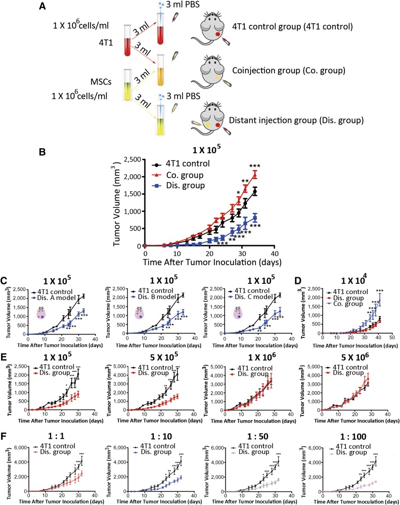
: Mesenchymal stem cells (MSCs) usually promote tumor growth and metastasis. By using a breast tumor 4T1 cell-based animal model, this study determined that coinjection and distant injection of allogeneic bone marrow-derived MSCs with tumor cells could exert different effects on tumor growth. Whereas the coinjection of MSCs with 4T1 cells promoted tumor growth, surprisingly, the injection of MSCs at a site distant from the 4T1 cell inoculation site suppressed tumor growth. We further observed that, in the distant injection model, MSCs decreased the accumulation of myeloid-derived suppressor cells and regulatory T cells in tumor tissues by enhancing proinflammatory factors such as interferon-γ, tumor necrosis factor-α, Toll-like receptor (TLR)-3, and TLR-4, promoting host antitumor immunity and inhibiting tumor growth. Unlike previous reports, this is the first study reporting that MSCs may exert opposite roles on tumor growth in the same animal model by modulating the host immune system, which may shed light on the potential application of MSCs as vehicles for tumor therapy and other clinical applications.

Significance: Mesenchymal stem cells (MSCs) have been widely investigated for their potential roles in tissue engineering, autoimmune diseases, and tumor therapeutics. This study explored the impact of coinjection and distant injection of allogeneic bone marrow-derived MSCs on mouse 4T1 breast cancer cells. The results showed that the coinjection of MSCs and 4T1 cells promoted tumor growth. MSCs might act as the tumor stromal precursors and cause immunosuppression to protect tumor cells from immunosurveillance, which subsequently facilitated tumor metastasis. Interestingly, the distant injection of MSCs and 4T1 cells suppressed tumor growth. Together, the results of this study revealed the dual functions of MSCs in immunoregulation.

Keywords: Breast tumor; Coinjection; Distant injection; Immunomodulatory; Mesenchymal stem cell.


Figures

Figure 1.
Figure 1.
Characteristics of MSCs and their effects on 4T1 cell proliferation, migration, invasion, and apoptosis. (A): GFP-4T1 cells were established to distinguish them from MSCs. (B): MSCs can differentiate into adipocytes, osteoblasts, and chondrocytes. (C): Surface markers of MSCs were detected by flow cytometry. (D): Proliferation curves of 4T1 cells, 4T1 cells cultured with MSC supernatant, coculture group, and control group. The doubling times of these groups were calculated. (E): Influence of MSCs on the invasion/migration ability of 4T1 cells. Numbers of migratory or invasive cells were counted in five random fields. (F): Effect of MSCs on GFP-4T1 cell apoptosis tested by Annexin V-PE and 7AAD staining. Apoptotic cell count was obtained after culture of GFP-4T1 cells with MSC supernatant and coculture with MSCs. Scale bars = 50 μm for all photographs. ∗∗∗, p < .001. Abbreviations: GFP, green fluorescent protein; MSC, mesenchymal stem cell; PE, phycoerythrin.
Figure 2.
Figure 2.
4T1 cell tumor growth is promoted by coinjection of mesenchymal stem cells (MSCs) but inhibited by distant injection of MSCs. (A): Strategy used to establish the experimental animal models. (B): Tumor volume was measured three times per week (n = 8; two-way analysis of variance [ANOVA]). (C): Changing the distant injection site still inhibited tumor growth (n = 5, two-way ANOVA). (D): An injection dose of 1 × 10⁴ MSCs or 4T1 cells per mouse was used, and tumor volume was measured (n = 5, two-way ANOVA). (E): Changing the injection dose influenced the efficiency of tumor growth inhibition (n = 5, two-way ANOVA). (F): Increasing the injection ratio amplified the tumor growth inhibition caused by distant injection (n = 5, two-way ANOVA). ∗, p < .05, ∗∗, p < .01, ∗∗∗, p < .001. Abbreviations: Co., coinjection; Dis., distant injection; PBS, phosphate-buffered saline.
Figure 3.
Figure 3.
Tumor growth promotion by coinjection was related to metastasis and the establishment of tumor stroma. Tumor growth inhibition caused by distant injection of MSCs may be related to angiogenesis. (A): Tumor tissues were collected on day 30 after tumor cell inoculation. Paraffin sections of the tumor samples were prepared to detect cellular morphology using H&E staining; Ki-67 and CD31 expression were examined using an IHC assay; and apoptotic status was evaluated using a TUNEL assay. (B): Primary lung cells were collected on day 7 after tumor cell inoculation. An ex vivo assay of primary culture cells from micrometastases was performed to assess lung metastasis during the early stage. 4T1 cells were cultured as a control and were stained using the Giemsa kit. (C): H&E staining results of lung metastases from different groups from the late stage (day 35 after tumor cell inoculation). The results were observed under ×200 and ×400 magnification. (D): Western blotting results for the epithelial-to-mesenchymal transition-related protein E-cadherin, vimentin, and tumor-associated fibroblast protein α-SMA. Tumor tissue proteins were extracted from the three groups. Three mice were used in each experiment: no. 1, 2, and 3 from the 4T1 control group; no. 4, 5, and 6 from the Co. group; and no. 7, 8, and 9 from the Dis. group. Scale bars = 50 μm for all photographs. Abbreviations: Co., coinjection; Dis., distant injection; HE, hematoxylin & eosin; IHC, immunohistochemistry; MSC, mesenchymal stem cell; SMA, smooth muscle actin TUNEL, transferase-mediated dUTP nick-end labeling.
Figure 4.
Figure 4.
Distant injection of 4T1 cells and mesenchymal stem cells (MSCs) before tumor cell inoculation inhibited tumor growth. Distant injection of 4T1 cells and MSCs also inhibited tumor growth when using CT26 mouse colorectal cancer cells. Additionally, immune T cells participated in the modulation of tumor growth caused by distant injection. (A): Distant injection of MSCs on day 3 before tumor cell inoculation inhibited tumor growth (n = 5, two-way analysis of variance [ANOVA]). (B): Tumor volume after inoculation with CT26 cells (n = 5, two-way ANOVA). (C): Effect of MSCs on tumor growth in BALB/c nude mice inoculated with 4T1 cells (n = 6, two-way ANOVA). (D): Adoptive transfer assay in BALB/c nude mice using 4T1 cells (n = 5, two-way ANOVA). (E): Relative tumor volume between control and the BALB/c nude mice subjected to adoptive transfer. Tumor growth in the 4T1 control group was set to 1. (F): Effect of MSCs on BCap37 human breast cancer cells in BALB/c nude mice (n = 5, two-way ANOVA). (G): Adoptive transfer assay in BALB/c nude mice using BCap37 cells (n = 5, two-way ANOVA). (H): Relative volume between control and BALB/c nude mice subjected to adoptive transfer. ∗∗, p < .01, ∗∗∗, p < .001. Abbreviations: AT, adoptive transfer; C, control; Co., coinjection; Dis., distant injection; RI, rate of inhibition.
Figure 5.
Figure 5.
Flow cytometry revealed changes in the proportion of immune cells in the spleens on day 30 after tumor cell inoculation. (A): Change in the proportion of Tregs (CD4+CD25+Foxp3+) of CD4+ T cells when gated for positive CD4-FITC cells (n = 3; one-way analysis of variance [ANOVA]). Proportions of MDSCs (CD11b+Gr-1+; n = 3; one-way ANOVA) (B), CD8+ T cells (n = 3; one-way ANOVA) (C), and APCs (CD80+MHC II+; n = 3; one-way ANOVA) (D) were also altered. ∗, p < .05, ∗∗∗, p < .001. Abbreviations: APC, antigen-presenting cells; Co., coinjection; Dis., distant injection; FITC, fluorescein isothiocyanate; MDSC, myeloid-derived suppressor cells; MHC, major histocompatibility complex; PE, phycoerythrin.
Figure 6.
Figure 6.
Quantitative polymerase chain reaction analysis of TNF-α, IFN-γ, TLR-3, TLR-4, inducible nitric oxide synthase, arginase, IL-1α, IL-1β, IL-6, and IL-12 mRNA expression in spleen cells at 12 hours, 24 hours, 3 days, 7 days, 20 days, and 30 days after tumor inoculation, presented as RQ compared with the expression in spleen cells from the 4T1 control group, which was set to 1. Data from three experiments are presented (n = 3; one-way ANOVA). ∗, p < .05, ∗∗, p < .01, ∗∗∗, p < .001. Abbreviations: ANOVA, analysis of variance; ARG, arginase; Co., coinjection; Dis., distant injection; IFN, interferon; IL, interleukin; iNOS, inducible nitric oxide synthase; RQ, relative quantification; TLR, toll-like receptor; TNF, tumor necrosis factor.
Figure 7.
Figure 7.
Cytokine, chemokine, and growth factor levels measured by enzyme-linked immunosorbent assay (ELISA) at 12 hours, 24 hours, 3 days, 5 days, 7 days, and 20 days after tumor cell inoculation. Peripheral blood was collected from the eye. Blood was placed for 20 minutes at room temperature, and the serum was collected after centrifugation. ELISA was carried out to detect TGF-β, TNF-α, IFN-γ, CXCL1, IL-10, and IL-21. Values are mean ± SEM (n = 3; one-way analysis of variance). ∗, p < .05, ∗∗, p < .01, ∗∗∗, p < .001. Abbreviations: Co., coinjection; CXCL, chemokine (C-X-C motif) ligand; Dis., distant injection; IFN, interferon; IL, interleukin; TGF, tumor growth factor; TNF, tumor necrosis factor.

References

    1. Keating A. Mesenchymal stromal cells: New directions. Cell Stem Cell. 2012;10:709–716. - PubMed
    1. Pittenger MF, Mackay AM, Beck SC, et al. Multilineage potential of adult human mesenchymal stem cells. Science. 1999;284:143–147. - PubMed
    1. Uchibori R, Tsukahara T, Mizuguchi H, et al. NF-κB activity regulates mesenchymal stem cell accumulation at tumor sites. Cancer Res. 2013;73:364–372. - PubMed
    1. Erices A, Conget P, Minguell JJ. Mesenchymal progenitor cells in human umbilical cord blood. Br J Haematol. 2000;109:235–242. - PubMed
    1. Baddoo M, Hill K, Wilkinson R, et al. Characterization of mesenchymal stem cells isolated from murine bone marrow by negative selection. J Cell Biochem. 2003;89:1235–1249. - PubMed

Publication types