. 2016 Jul 21;61(14):5253-74.
doi: 10.1088/0031-9155/61/14/5253. Epub 2016 Jun 29.

Efficacy of histotripsy combined with rt-PA in vitro

Affiliations

Efficacy of histotripsy combined with rt-PA in vitro

Kenneth B Bader et al. Phys Med Biol. .

Abstract

Histotripsy, a form of therapeutic ultrasound that uses the mechanical action of microbubble clouds for tissue ablation, is under development to treat chronic deep vein thrombosis (DVT). We hypothesize that combining thrombolytic agents with histotripsy will enhance clot lysis. Recombinant tissue plasminogen activator (rt-PA) and rt-PA-loaded echogenic liposomes that entrain octafluoropropane microbubbles (OFP t-ELIP) were used in combination with highly shocked histotripsy pulses. Fully retracted porcine venous clots, with similar features of DVT occlusions, were exposed either to histotripsy pulses alone (peak negative pressures of 7-20 MPa), histotripsy and OFP t-ELIP, or histotripsy and rt-PA. Microbubble cloud activity was monitored with passive cavitation imaging during histotripsy exposure. The power levels of cavitation emissions from within the clot were not statistically different between treatment types, likely due to the near instantaneous rupture and destruction of OFP t-ELIP. The thrombolytic efficacy was significantly improved in the presence of rt-PA. These results suggest the combination of histotripsy and rt-PA could serve as a potent therapeutic strategy for the treatment of DVT.
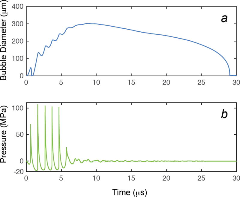

Figures

Fig 1
Fig 1
Overhead view of experimental set up for histotripsy ablation of porcine clots. The L7-4 imaging array was oriented to monitor cavitation activity along the axial/elevational plane of the histotripsy transducer. The elevational dimension, not shown, is perpendicular to both the lateral and axial dimensions (into the page).
Fig 2
Fig 2
Representative scattered power spectra acquired from acoustic emissions generated by histotripsy-induced microbubble clouds within a porcine whole blood clot without OFP t-ELIP (a) and with OFP t-ELIP (b) (1-MHz fundamental frequency, 5 µs pulse duration, 20 Hz pulse repetition frequency). The peak negative pressures for each insonation are indicated in the legend. Each power spectra was normalized by the maximum power while insonifying with a peak negative pressure of 20 MPa either without (a) or with (b) OFP t-ELIP.
Fig 2
Fig 2
Representative scattered power spectra acquired from acoustic emissions generated by histotripsy-induced microbubble clouds within a porcine whole blood clot without OFP t-ELIP (a) and with OFP t-ELIP (b) (1-MHz fundamental frequency, 5 µs pulse duration, 20 Hz pulse repetition frequency). The peak negative pressures for each insonation are indicated in the legend. Each power spectra was normalized by the maximum power while insonifying with a peak negative pressure of 20 MPa either without (a) or with (b) OFP t-ELIP.
Fig 3
Fig 3
(Top panel) For a given data set, all 400 PCI frames were temporally averaged (hot colormap). The axial and elevational directions are in reference to the histotripsy transducer (Fig. 1). The histotripsy pulse is propagating along the axial dimension (from left to right) in the image. The artifact along the elevational direction of the histotripsy transducer occurs because of the elongated point spread function in the direction normal to the face of the linear array (Haworth et al 2012). A duplex averaged PCI/plane wave B-mode image (grayscale) allowed comparison of the temporally averaged acoustic power with the clot. The dotted and dashed line notes the elevational position of the maximum pixel amplitude of the PCI. (Bottom panel) The temporally averaged PCI amplitude as a function of the axial dimension at a fixed elevational location (dotted and dashed line in top panel). The boundaries of the lumen are denoted in green, and the clot boundaries are noted in blue.
Fig 4
Fig 4
a. The mean volume-weighted size distribution measurements of OFP t-ELIP between 0.65 and 10 µm. The error bars represent one standard deviation (N = 5). b. The measured attenuation coefficient as a function of frequency (solid line) and theoretical fit to the de Jong model (dashed line) based on the estimated shell parameters of OFP t-ELIP. The error bars represent one standard deviation (N = 5). The coefficient of determination of the fit to the data was 0.91. The number density of liposomes employed for the attenuation measurement was 1.9 ×10⁸ liposomes/mL. Following Raymond et al (2014), the estimated number density of bubbles for the attenuation measurement was 5.4 ×10⁶ bubbles/mL.
Fig 4
Fig 4
a. The mean volume-weighted size distribution measurements of OFP t-ELIP between 0.65 and 10 µm. The error bars represent one standard deviation (N = 5). b. The measured attenuation coefficient as a function of frequency (solid line) and theoretical fit to the de Jong model (dashed line) based on the estimated shell parameters of OFP t-ELIP. The error bars represent one standard deviation (N = 5). The coefficient of determination of the fit to the data was 0.91. The number density of liposomes employed for the attenuation measurement was 1.9 ×10⁸ liposomes/mL. Following Raymond et al (2014), the estimated number density of bubbles for the attenuation measurement was 5.4 ×10⁶ bubbles/mL.
Fig 5
Fig 5
Thrombolytic efficacy for histotripsy alone, histotripsy and OFP t-ELIP, and histotripsy and rt-PA reported in terms of percent clot mass loss. The p values between treatment arms are indicated in Table 1. The asterisks (*) indicate that the thrombolytic efficacy of that treatment arm is greater than all other treatment arms for that particular peak negative pressure (p < 0.05). The error bars represent one standard deviation (N = 6).
Fig 6
Fig 6
Size distribution of debris measured after treatment at 0 MPa (a), 7 MPa (b), 14 MPa (c), and 20 MPa (d) peak negative pressure. The error bars represent one standard deviation (N = 3). Note that residual lipid and rt-PA may be present in addition to clot debris for the rt-PA and OFP t-ELIP arms.
Fig 6
Fig 6
Size distribution of debris measured after treatment at 0 MPa (a), 7 MPa (b), 14 MPa (c), and 20 MPa (d) peak negative pressure. The error bars represent one standard deviation (N = 3). Note that residual lipid and rt-PA may be present in addition to clot debris for the rt-PA and OFP t-ELIP arms.
Fig 7
Fig 7
The acoustic power within the clot for each treatment arm. The error bars represent one standard deviation (N = 6). The acoustic power within the clot significantly increased with the peak negative pressure of the histotripsy pulse for all treatment arms. At a given peak negative pressure, there was no significant difference between the treatment arms.
Fig 8
Fig 8
The variability of the location of the maximum PCI pixel amplitude along the axial dimension of the histotripsy source as a function of the peak negative pressure of the histotripsy pulse. The error bars represent one standard deviation (N = 6). The variability significantly decreases with the peak negative pressure for histotripsy and histotripsy and OFP t-ELIP. The asterisks (*) indicate that the axial variability of that treatment arm is greater than all other treatment arms for that particular peak negative pressure of the histotripsy pulse.
Fig 9
Fig 9
The response of a shelled microbubble using the Marmottant equation (Strandness and Van Breda 1994, Marmottant et al 2005b), Eq. (1), is shown in panel a. The viscoelastic shell properties of OFP t-ELIP derived from the attenuation measurements were used in the calculation. The experimentally measured waveform used as the excitation source in the computation is shown in panel b. The initial diameter of the cavitation nuclei was 1.7 µm.
Fig 10
Fig 10
Maximum size of the bubble as a function of initial size for shelled microbubbles calculated by numerical integration of the Marmottant model (symbols) and unshelled microbubbles without shell encapsulation using the analytic theory of Bader and Holland (solid lines). The peak negative pressure of the histotripsy pulse is shown in the legend.
Fig 11
Fig 11
Percent of debris measured after treatment between 4–5 µm, the typical size of porcine red blood cells, compared to the total number of particles measured (0.65–18 µm). The error bars represent one standard deviation (N = 3). The addition of thrombolytic significantly increased the number of particulates in this range, suggesting preservation of red blood cells for the combined histotripsy and thrombolytic treatment arms.

References

    1. Arslan B, Turba U, Matsumoto A. Acute Renal Failure Associated with Percutaneous Mechanical Thrombectomy for Iliocaval Venous Thrombosis. Semin intervent Radiol. 2007;24:288–95. - PMC - PubMed
    1. Augustinos P. Invasive Approaches to Treatment of Venous Thromboembolism. Circulation. 2004;110:I-27–I-34. - PubMed
    1. Avgerinos ED, Hager ES, Naddaf A, Dillavou E, Singh M, Chaer RA. Outcomes and predictors of failure of thrombolysisfor iliofemoral deep venous thrombosis. J Vasc Surg. 2015;3:35–41. - PubMed
    1. Aziz F, Comerota AJ. Quantity of residual thrombus after successful catheter-directed thrombolysis for iliofemoral deep vein thrombosis correlates with recurrence. Eur J Vasc Endovasc Surg. 2012;44:210–3. - PubMed
    1. Bader KB, Holland CK. Predicting the growth of nanoscale nuclei by histotripsy pulses. Phys Med Biol. 2016 (In Press) - PMC - PubMed