. 2016 Dec 1;594(23):6817-6831.
doi: 10.1113/JP272633. Epub 2016 Aug 29.

Modular modelling with Physiome standards

Affiliations

Modular modelling with Physiome standards

Michael T Cooling et al. J Physiol. .

Abstract

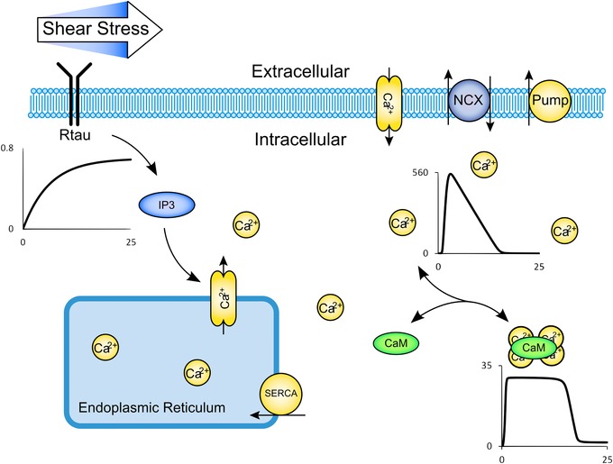
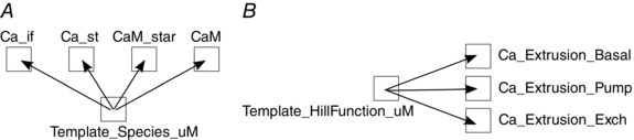
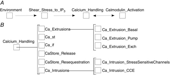
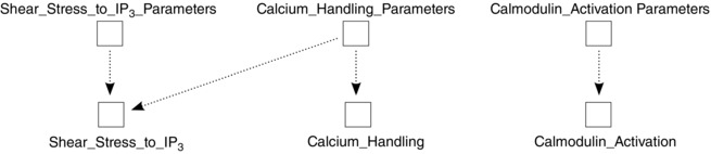
Key points: The complexity of computational models is increasing, supported by research in modelling tools and frameworks. But relatively little thought has gone into design principles for complex models. We propose a set of design principles for complex model construction with the Physiome standard modelling protocol CellML. By following the principles, models are generated that are extensible and are themselves suitable for reuse in larger models of increasing complexity. We illustrate these principles with examples including an architectural prototype linking, for the first time, electrophysiology, thermodynamically compliant metabolism, signal transduction, gene regulation and synthetic biology. The design principles complement other Physiome research projects, facilitating the application of virtual experiment protocols and model analysis techniques to assist the modelling community in creating libraries of composable, characterised and simulatable quantitative descriptions of physiology.

Abstract: The ability to produce and customise complex computational models has great potential to have a positive impact on human health. As the field develops towards whole-cell models and linking such models in multi-scale frameworks to encompass tissue, organ, or organism levels, reuse of previous modelling efforts will become increasingly necessary. Any modelling group wishing to reuse existing computational models as modules for their own work faces many challenges in the context of construction, storage, retrieval, documentation and analysis of such modules. Physiome standards, frameworks and tools seek to address several of these challenges, especially for models expressed in the modular protocol CellML. Aside from providing a general ability to produce modules, there has been relatively little research work on architectural principles of CellML models that will enable reuse at larger scales. To complement and support the existing tools and frameworks, we develop a set of principles to address this consideration. The principles are illustrated with examples that couple electrophysiology, signalling, metabolism, gene regulation and synthetic biology, together forming an architectural prototype for whole-cell modelling (including human intervention) in CellML. Such models illustrate how testable units of quantitative biophysical simulation can be constructed. Finally, future relationships between modular models so constructed and Physiome frameworks and tools are discussed, with particular reference to how such frameworks and tools can in turn be extended to complement and gain more benefit from the results of applying the principles.

Keywords: modelling; modularity; physiome; standards.


Figures

Figure 1
Figure 1. Biological schematic diagram of the Signalling Model Example
Shear stress sensed by receptors (Rtau) leads to IP3 production and store calcium release. Calcium is also transferred between the extra‐ and intracellular compartments via stress‐sensitive calcium channels, calcium/sodium exchanger, a calcium pump, a basal calcium leak and capacitative calcium entry. Calcium leads to activation of calmodulin (CaM). Representative traces of Rtau, free intracellular calcium, and activated calmodulin over a 25 s simulated time period are shown (please see the Supporting information for more details on the traces).
Figure 2
Figure 2. Common template components being imported to form biological entities (A) and processes (B)
Squares are CellML components, and arrows show the direction of the import: for example, ‘Ca_if’ is an imported instance of ‘Template_Species_uM’. Ca_if and Ca_st are free and store calcium respectively, ‘CaM’ is calmodulin, and the ‘_star’ suffix denotes the activated version.
Figure 3
Figure 3. The biological component hierarchy of the Signalling Example model
A, the top level of the hierarchy, with large cohesive modules (dotted lines indicate general flow of information; free intracellular calcium modulates both IP3 generation and calmodulin activation). B, the nested nature (brackets indicate encapsulation relationships) of the Calcium_Handling module, which are constructed from smaller cohesive components representing sub‐modules. Similar sub‐hierarchies could be drawn for Shear_Stress_to_IP3 and Calmodulin_Activation components.
Figure 4
Figure 4. Variables of the ‘Ca_st_delta’ component, which handles multi‐module communication between Ca_st (store calcium) and other components, with respect to flux
Arrows represent input or output on variables listed by name. The component includes ‘input’ variables for fluxes (positive or negative) from store resequestration and release processes, as well as any external processes (see the example models for more details). There is also a variable for converting between different volumes (in this case, cytoplasm/store). The net flux is component output, named ‘JNet’. The ‘Ca_st’ component will use ‘JNet’ to update the total amount of calcium in the store, across a given time interval.
Figure 5
Figure 5. Parameters and initial conditions are placed in their own components, here for each high level biological component
Each has their own parameter set, but the separation allows for parameters to come from other sources too. Shear_Stress_to_IP3 can run using its own parameters; however, when linked with other modules (such as Calcium_Handling) it may be more appropriate for some parameters to come from that source instead. Following Principle (6) allows such ‘parameter collisions’ to be resolved by the model builder.
Figure 6
Figure 6. Units reuse
Units (triangles) are defined at the lowest level and imported into the models (circles) housing low‐level components so that those components can be reused in other models along with consistent and expected unit information (continuous arrows show unit import directions). Units for higher level modules (‘Calcium_Handling’, in this case) may be (dotted arrows) imported from lower levels or from other ‘child’ models. Additionally, following Principle (8), we include a ‘Units_’ model that houses standard units providing a library of such units for many models to use promoting inter‐model consistency. Higher level constructs can import all units directly from that common source, reducing coupling that the former strategy would incur.
Figure 7
Figure 7. Biological schematic diagram of the Core Domains model
The membrane is depolarised at t = 50s (voltage trace shown, scale is mV), resulting in calcium influx (trace shown in μm) through L‐type calcium channels (electrophysiology). The channels are sensitive to pH and [ATP], both of which are influenced by myosin ATPase reaction (metabolism). Calcium activates calmodulin and calcineurin (trace of ratio of active calcineurin–calmodulin complex shown), leading to NFAT cycling (signalling). NFAT (trace shown in nm) is a transcription factor for a sample ‘device’ (gene regulation and synthetic biology) resulting in mRNA production that is translated outside the nucleus to form a GFP signal (trace shown in nm), which increases as the membrane is depolarised. Please see the Supporting information for more details.
Figure 8
Figure 8. Simultaneous model reuse and characterisation
A, model aggregation and re‐parameterisation. Here all models (rounded boxes) are complete and simulatable in their own rights. Components (squares) imported (dotted arrows) from ‘NFAT_Cycling’ and ‘Calcineurin_Activation’ form part of the higher level model ‘CaN_to_DNAReady_NFAT’, which includes parameter components related to the child components by default, but could equally source parameter values from other components. ‘Calcium_to_Calcineurin_Activation’ model imports the ‘Calcineurin_Activation components’, but also includes a calcium species whose concentration varies over time, hence replaces the constant calcium concentration parameter with the output of the ‘Ca’ component. B, output of the ’Calcium_to_Calcineurin’ model. The trace of activated calcineurin proportion for increasing calcium concentration is shown (calcineurin and calmodulin concentrations are fixed in that model).

References

    1. Bartholet RG, Brogan DC, Reynolds PF & Carnahan JC (2004). In search of the philosopher's stone: Simulation composability versus component‐based software design. Proceedings of the 2004 Fall Simulation Interoperability Workshop, Orlando, FL, September 2004 Simulation Interoperability Standards Organization, Orlando, FL, USA.
    1. Beard DA, Neal ML, Tabesh‐Saleki N, Thompson CT, Bassingthwaighte JB, Shimoyama M & Carlson BE (2012). Multiscale modeling and data integration in the virtual physiological rat project. Ann Biomed Eng 40, 2365–2378. - PMC - PubMed
    1. Büchel F, Rodriguez N, Swainston N, Wrzodek C, Czauderna T, Keller R, Mittag F, Schubert, M , Glont M, Golebiewski M, van Iersel M, Keating S, Rall M, Wybrow M, Hermjakob H, Hucka M, Kell DB, Muller W, Mendes P, Zell A, Chaouiya C, Saez‐Rodriguez J, Schreiber F, Laibe C, Dräger A & Le Novère N (2013). Path2Models: Large‐scale generation of computational models from biochemical pathway maps. BMC Syst Biol 7, 116. - PMC - PubMed
    1. Cooling M, Hunter P & Crampin EJ (2007). Modeling hypertrophic IP3 transients in the cardiac myocyte. Biophys J 93, 3421–3433. - PMC - PubMed
    1. Cooling MT, Hunter P & Crampin EJ (2009). Sensitivity of NFAT cycling to cytosolic calcium concentration: implications for hypertrophic signals in cardiac myocytes. Biophys J 96, 2095–2104. - PMC - PubMed

Publication types

LinkOut - more resources