. 2017 Mar;10(2):434-445.
doi: 10.1038/mi.2016.55. Epub 2016 Jun 29.

NLRP6 function in inflammatory monocytes reduces susceptibility to chemically induced intestinal injury

Affiliations

NLRP6 function in inflammatory monocytes reduces susceptibility to chemically induced intestinal injury

S S Seregin et al. Mucosal Immunol. 2017 Mar.

Abstract

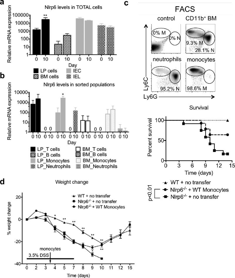
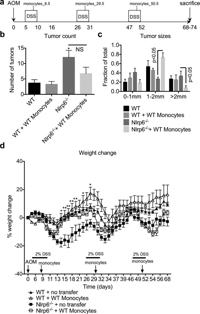
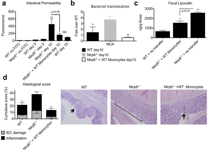
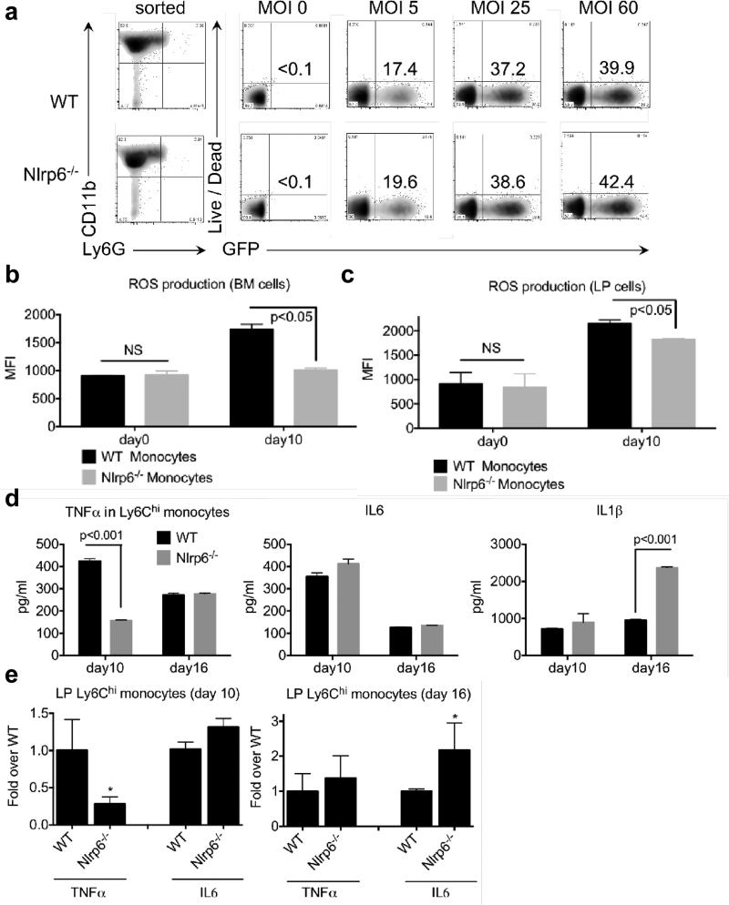
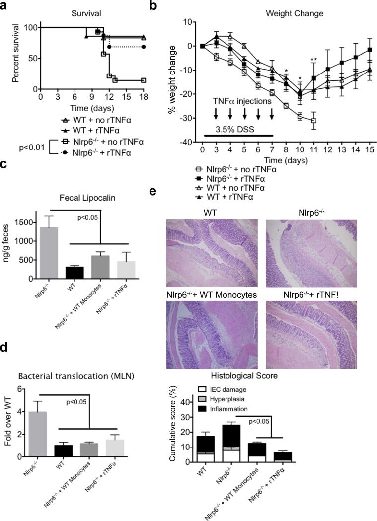
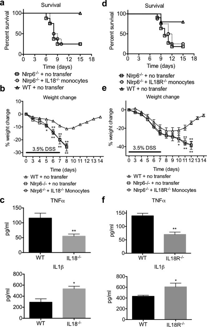
NLRP6 is a member of the Nod-like receptor family, whose members are involved in the recognition of microbes and/or tissue injury. NLRP6 was previously demonstrated to regulate the production of interleukin (IL)-18 and is important for protecting mice against chemically induced intestinal injury and colitis-associated colon cancer. However, the cellular mechanisms by which NLRP6 reduces susceptibility to colonic inflammation remain unclear. Here, we determined that NLRP6 expression is specifically upregulated in Ly6Chi inflammatory monocytes that infiltrate into the colon during dextran sulfate sodium (DSS)-induced inflammation. Adoptive transfer of wild-type (WT) Ly6Chi inflammatory monocytes into Nlrp6-/- mice was sufficient to protect them from mortality, significantly reducing intestinal permeability and damage. NLRP6-deficient inflammatory monocytes were defective in tumor necrosis factor α (TNFα) production, which was important for reducing DSS-induced mortality and was dependent on autocrine IL-18 signaling by inflammatory monocytes. Our data reveal a previously unappreciated role for NLRP6 in inflammatory monocytes, which are recruited after DSS-induced intestinal injury to promote barrier function and limit bacteria-driven inflammation. This study highlights the importance of early cytokine responses, particularly NLRP6-dependent and IL-18-dependent TNFα production, in preventing chronic dysregulated inflammation.


Figures

Figure 1
Figure 1. NLRP6 is induced in lamina propria Ly6Chi monocytes during DSS induced inflammation, and is important for reducing susceptibility to colitis
(a) NLRP6 levels were measured in BM, LP, IECs and IELs of WT mice at day 0 and day 10 of AOM/DSS by qPCR. (b) NLRP6 expression was measured in CD3+B220 CD11b T cells, CD3 B220+CD11b B cells, CD3 CD11b+Ly6ChiLy6G monocytes and CD3 CD11b+Ly6CintLy6G+ neutrophils within the BM and LP. Data are representative of three independent experiments; n=11 for day 0, n=10 for day 10. *, ** - p<0.05, p<0.001, respectively, as compared to day 0 time point of the same genotype. (c) Representative plots of Ly6C versus Ly6G staining of CD11b+ BM cells (top). Kaplan–Meyer survival curves of mice treated with 7 days of 3.5% DSS (bottom). (d) Percent weight change with 3.5% DSS administration. Data are representative of two independent experiments; n=15, n=24, n=14 for WT, Nlrp6−/− and Nlrp6−/− + WT Ly6Chi monocytes groups respectively. *, ** - p<0.05, p<0.001, respectively, as compared to Nlrp6−/−.
Figure 2
Figure 2. Adoptive transfer of WT Ly6Chi monocytes into Nlrp6−/− mice significantly reduces tumor growth in AOM/DSS model of colitis-associated tumorigenesis
(a) Age and sex-matched Nlrp6−/− and WT mice were treated with AOM followed by three 5-day cycles of 2% DSS. Sorted Ly6Chi monocytes were injected i.v. 3.5 days after the start of each DSS cycle. Three weeks after the last cycle of DSS mice were sacrificed and tumors grossly counted with a stereomicroscope. Number (b) and size (c) of tumors are shown. (d) Percent weight change during AOM/DSS treatment. Data are representative of three independent experiments, n=15, n=10, n=19, n=22 for WT, WT + WT Ly6Chi monocytes, Nlrp6−/− and Nlrp6−/− + WT Ly6Chi monocytes groups respectively. *, ** - p<0.05, p<0.001, respectively, as compared to WT (b) or Nlrp6−/− mice (d).
Figure 3
Figure 3. Adoptive transfer of WT Ly6Chi monocytes into Nlrp6−/− mice limits bacterial translocation and intestinal damage
Age- and sex-matched Nlrp6−/− and WT mice were treated with AOM followed by 5 days of 2% DSS. WT Ly6Chi monocytes were adoptively transferred into Nlrp6−/− mice 3.5 days after start of DSS. (a) Mice were gavaged FITC-dextran at end of 5 days of DSS followed by serum collection and measurement of fluorescence 4 hours later. (b) Normalized levels of total bacteria/MLN as measured by qPCR after 5 days of 2% DSS. (c) Fecal lipocalin-2 levels as measured by ELISA after 5 days of DSS. (d) Histological inflammatory scores based on extent of inflammatory cell infiltration and IEC damage;Micrographs of H&E sections are at 200x magnification. Arrow points to focal erosion with inflammatory infiltrate. Bracket indicates ulcerated epithelium with inflammatory infiltrate in LP and submucosa. Data are representative of three independent experiments, n=15, n=16, n=20 for WT, Nlrp6−/− and Nlrp6−/− + WT Ly6Chi monocytes groups respectively. *, ** - p<0.05, p<0.001, respectively, as compared to day 0 of both genotypes (a), or as compared to Nlrp6−/− (b,c) or as compared to WT (d).
Figure 4
Figure 4. NLRP6 deficient Ly6Chi monocytes have reduced ROS and TNFα production during the acute inflammatory response to DSS
(a) Ly6Chi monocytes were sorted from WT or Nlrp6−/− mice and incubated with GFP-labeled E.coli at MOI 0, 5, 25 and 60. Phagocytosis was measured by percent GFP+ Ly6Chi monocytes. Data are representative of two independent experiments, n=6. ROS production (MFI) as measured by CellROX deep red in BM- (b) or LP-derived (c) Ly6Chi monocytes at the indicated time points. Ly6Chi monocytes from WT or Nlrp6−/− mice were sorted from the LP after 5 days of 2% DSS (day 10, AOM/DSS model) or 6 days after completion of DSS (day 16, AOM/DSS model) and cytokine levels were measured by ELISA (d) and by qPCR (e). Data are representative of at least three independent experiments, mean ± SEM; n=14/genotype, (day 10); n=12/genotype (day 0); n=8/genotype (day 16). * - p<0.05, as compared to WT.
Figure 5
Figure 5. Recombinant TNFα administration into Nlrp6−/− mice early during DSS treatment reduces susceptibility to DSS-induced colitis
(a) Kaplan–Meyer survival curves and (b) percent weight change of age- and sex-matched WT and Nlrp6−/− mice treated with 3.5% DSS for 7 days with or without rTNFα injections given on days 3-7. Data are representative of two independent experiments, mean ± SEM; n=15, n=16 for Nlrp6−/− and Nlrp6−/− + 1 μg/mouse rTNFα respectively; n=5 for WT and WT + 1 μg/mouse rTNFα (c) Fecal lipocalin-2 levels measured in WT and Nlrp6−/− mice (day 10, AOM/DSS model) after adoptive transfer of Ly6Chi monocytes or 1 μg TNF given on days 3-5 of DSS (d) Normalized levels of total bacteria/MLN as measured by qPCR after 5 days of 2% DSS (day 10 AOM/DSS model). (e) H&E micrographs at 100x after 5 days of 2% DSS (top) with histological scoring are shown. Bracket indicates diffuse ulceration with inflammatory infiltrate extending throughout the LP and submucosa; n=5, n=6, n=6, n=7 for WT, Nlrp6−/−, Nlrp6−/− + 1 μg/mouse rTNFα, and Nlrp6−/− + WT Ly6Chi monocytes groups respectively.
Figure 6
Figure 6. Autocrine IL-18 production by Ly6Chi monocytes protects against DSS-induced colitis by increasing TNFα production
Ly6Chi monocytes were purified from WT, Il18−/− or Il18r1−/− mice and i.v. injected (2×10⁶ cells/mouse) into Nlrp6−/− recipients at day 3.5 of a 7 day course of 3.5% DSS. Mice survival (a) and weight loss (b) for WT, Nlrp6−/− and Nlrp6−/− + Il18−/− Ly6Chi monocytes was assessed. n=8/group (c) Ly6Chi monocytes were sorted from the LP after 5 days of 2% DSS (day 10 AOM/DSS model) from Il18−/− mice, and cytokine levels in overnight cultures were measured by ELISA. n=4 per group. Mice survival (d) and weight loss (e) after adoptive transfer of Il18R−/− Ly6Chi monocytes into Nlpr6−/− mice. n=5 for WT, n=11 for Nlrp6−/− and n=8 for Nlrp6−/− + Il18r1−/− Ly6Chi monocytes (f) Cytokine levels measured from culture supernatants of WT and Il18R−/− Ly6Chi monocytes after 5 days of 2% DSS (day 10 AOM/DSS model), n=4/group. *, ** - p<0.05, p<0.001.

References

    1. Chen GY, Liu M, Wang F, Bertin J, Nunez G. A functional role for Nlrp6 in intestinal inflammation and tumorigenesis. J Immunol. 2011;186(12):7187–7194. - PMC - PubMed
    1. Chen GY. Role of Nlrp6 and Nlrp12 in the maintenance of intestinal homeostasis. Eur J Immunol. 2013;44(2):321–327. - PMC - PubMed
    1. Elinav E, Strowig T, Kau AL, Henao-Mejia J, Thaiss CA, Booth CJ, et al. NLRP6 inflammasome regulates colonic microbial ecology and risk for colitis. Cell. 2011;145(5):745–757. - PMC - PubMed
    1. Normand S, Delanoye-Crespin A, Bressenot A, Huot L, Grandjean T, Peyrin-Biroulet L, et al. Nod-like receptor pyrin domain-containing protein 6 (NLRP6) controls epithelial self-renewal and colorectal carcinogenesis upon injury. Proc Natl Acad Sci U S A. 2011;108(23):9601–9606. - PMC - PubMed
    1. Allen IC, TeKippe EM, Woodford RM, Uronis JM, Holl EK, Rogers AB, et al. The NLRP3 inflammasome functions as a negative regulator of tumorigenesis during colitis associated cancer. J Exp Med. 2010;207(5):1045–1056. - PMC - PubMed

Publication types

MeSH terms