. 2016 Aug 22;60(9):5357-67.
doi: 10.1128/AAC.01073-16. Print 2016 Sep.

In Vitro Assessment of Combinations of Enterovirus Inhibitors against Enterovirus 71

Affiliations

In Vitro Assessment of Combinations of Enterovirus Inhibitors against Enterovirus 71

Yizhuo Wang et al. Antimicrob Agents Chemother. .

Abstract

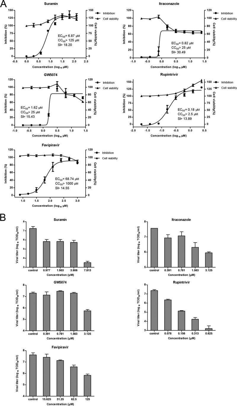
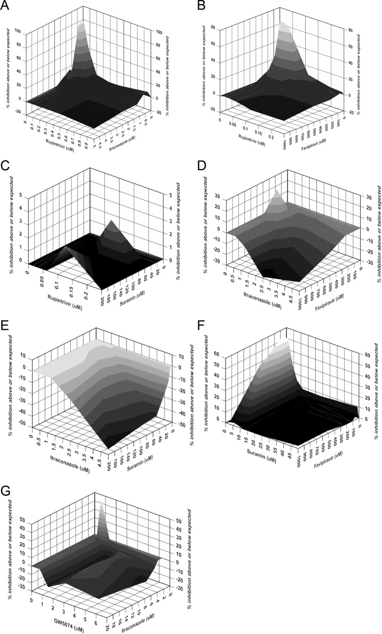
Enterovirus 71 (EV-A71) is a major causative pathogen of hand, foot, and mouth disease (HFMD) epidemics. No antiviral therapies are currently available for treating EV-A71 infections. Here, we selected five reported enterovirus inhibitors (suramin, itraconazole [ITZ], GW5074, rupintrivir, and favipiravir) with different mechanisms of action to test their abilities to inhibit EV-A71 replication alone and in combination. All selected compounds have anti-EV-A71 activities in cell culture. The combination of rupintrivir and ITZ or favipiravir was synergistic, while the combination of rupintrivir and suramin was additive. The combination of suramin and favipiravir exerted a strong synergistic antiviral effect. The observed synergy was not due to cytotoxicity, as there was no significant increase in cytotoxicity when compounds were used in combinations at the tested doses. To investigate the potential inhibitory mechanism of favipiravir against enterovirus, two favipiravir-resistant EV-A71 variants were independently selected, and both of them carried an S121N mutation in the finger subdomain of the 3D polymerase. Reverse engineering of this 3D S121N mutation into an infectious clone of EV-A71 confirmed the resistant phenotype. Moreover, viruses resistant to ITZ or favipiravir remained susceptible to other inhibitors. Most notably, combined with ITZ, rupintrivir prevented the development of ITZ-resistant variants. Taken together, these results provide a rational basis for the design of combination regimens for use in the treatment of EV-A71 infections.


Figures

FIG 1
FIG 1
Antiviral activities of individual compounds against EV-A71. (A) Antiviral activities and cytotoxicities of individual compounds. Twofold serial dilutions of compounds were added to RD cells, and the inhibitory effects of the compounds were analyzed by a CPE assay. EC50s were calculated by using Prism nonlinear regression (GraphPadPrism5). Cytotoxicity was also examined by incubation of RD cells with the indicated concentrations of compounds. Cell viability was measured by using CellTiter-Glo reagent and is presented as the percent luminescence derived from the compound-treated cells compared to that from the mock-treated cells. (B) Antiviral effect of selected compounds on virus yield. RD cells were infected with EV-A71 strain FY573 at an MOI of 0.1 and treated with 2-fold serial dilutions of compounds. Supernatants were collected at 48 h postinfection, and viral titers were determined by a TCID50 assay. The data shown were obtained from two independent replicates. Error bars indicate the standard deviations from two independent experiments.
FIG 2
FIG 2
Mutation in EV-A71 3D polymerase confers resistance to favipiravir. (A) Scheme for the selection of favipiravir-resistant virus. (B) Resistance analysis by a virus yield reduction assay. Vero cells were infected with the WT virus, P16 selections (Sel I and Sel III), or the 3D S121N mutant virus in the presence of 600 μM favipiravir or 0.25% DMSO (as a negative control). At 48 h postinfection, the viral titers in culture fluids were quantified by a plaque assay. Fold reduction was determined by dividing the titer of virus treated with 0.25% DMSO by that of virus treated with 600 μM favipiravir. Resistance is quantified by the fold change compared to the wild-type virus, which was set as 1.0. (C) Summary of mutations identified from the two selections. Locations of the nucleotide and/or amino acid changes are indicated. (D) Sequence alignment of the resistance regions in 3D proteins of representative strains of EV-A71 genotypes (left) (the name of each EV-A71 strain of genotypes A, B1, B2, B3, B4, B5, C1, C2, C3, C4, and C5 is indicated on the left) and members of the genus Enterovirus (right) (sequences with GenBank accession no. EU262658 [CV-A16], M33854 [CV-B3], KF537633 [PV1], and KF726085 [EV-D68] were used). The arrow indicates the mutation in favipiravir-resistant EV-A71 identified in this study. The secondary structure based on the structure model of the EV-A71 3D polymerase (Protein Data Bank accession no. 3N6L) is shown above the sequence. (E) Location of the S121 mutation in the structural model of EV-A71 3D polymerase. Residue S121 is shown in red. The figure was produced by using PyMOL. (F) Modeling of favipiravir into the finger subdomain of EV-A71 3D polymerase. (Left) Favipiravir (represented in stick form and colored in atoms) was docked into the structure of EV-A71 3Dpol (Protein Data Bank accession no. 3N6L) (gray) by using Autodock Vina, and the lowest-energy conformation is presented. Residue S121 is shown in red. (Right) Partial structure of the 3Dpol finger subdomain represented as a cartoon. Residue S121 is highlighted in a stick representation, with carbon atoms in green. The image was generated by using PyMOL. (G) Modeling of the favipiravir-resistant mutation. (Left) The polymerase residues in contact with favipiravir are shown with carbon atoms in green and explicitly labeled. (Right) Structural model of the 3D S121N mutation generated by using SWISS-MODEL. Favipiravir is shown in cyan, and both residues S121 and N121 are shown in magenta. Images were created by using PyMOL. (H) Phenotypic characterization of the resistance mutant. (Left) Growth kinetics of wild-type EV-A71 and the 3D S121N mutant virus. The data shown were obtained from two independent replicates. Error bars indicate the standard deviations from two independent experiments. (Right) Plaque phenotypes of wild-type EV-A71 and the 3D S121N mutant virus.
FIG 2
FIG 2
Mutation in EV-A71 3D polymerase confers resistance to favipiravir. (A) Scheme for the selection of favipiravir-resistant virus. (B) Resistance analysis by a virus yield reduction assay. Vero cells were infected with the WT virus, P16 selections (Sel I and Sel III), or the 3D S121N mutant virus in the presence of 600 μM favipiravir or 0.25% DMSO (as a negative control). At 48 h postinfection, the viral titers in culture fluids were quantified by a plaque assay. Fold reduction was determined by dividing the titer of virus treated with 0.25% DMSO by that of virus treated with 600 μM favipiravir. Resistance is quantified by the fold change compared to the wild-type virus, which was set as 1.0. (C) Summary of mutations identified from the two selections. Locations of the nucleotide and/or amino acid changes are indicated. (D) Sequence alignment of the resistance regions in 3D proteins of representative strains of EV-A71 genotypes (left) (the name of each EV-A71 strain of genotypes A, B1, B2, B3, B4, B5, C1, C2, C3, C4, and C5 is indicated on the left) and members of the genus Enterovirus (right) (sequences with GenBank accession no. EU262658 [CV-A16], M33854 [CV-B3], KF537633 [PV1], and KF726085 [EV-D68] were used). The arrow indicates the mutation in favipiravir-resistant EV-A71 identified in this study. The secondary structure based on the structure model of the EV-A71 3D polymerase (Protein Data Bank accession no. 3N6L) is shown above the sequence. (E) Location of the S121 mutation in the structural model of EV-A71 3D polymerase. Residue S121 is shown in red. The figure was produced by using PyMOL. (F) Modeling of favipiravir into the finger subdomain of EV-A71 3D polymerase. (Left) Favipiravir (represented in stick form and colored in atoms) was docked into the structure of EV-A71 3Dpol (Protein Data Bank accession no. 3N6L) (gray) by using Autodock Vina, and the lowest-energy conformation is presented. Residue S121 is shown in red. (Right) Partial structure of the 3Dpol finger subdomain represented as a cartoon. Residue S121 is highlighted in a stick representation, with carbon atoms in green. The image was generated by using PyMOL. (G) Modeling of the favipiravir-resistant mutation. (Left) The polymerase residues in contact with favipiravir are shown with carbon atoms in green and explicitly labeled. (Right) Structural model of the 3D S121N mutation generated by using SWISS-MODEL. Favipiravir is shown in cyan, and both residues S121 and N121 are shown in magenta. Images were created by using PyMOL. (H) Phenotypic characterization of the resistance mutant. (Left) Growth kinetics of wild-type EV-A71 and the 3D S121N mutant virus. The data shown were obtained from two independent replicates. Error bars indicate the standard deviations from two independent experiments. (Right) Plaque phenotypes of wild-type EV-A71 and the 3D S121N mutant virus.
FIG 3
FIG 3
Analysis of drug combinations using the MacSynergy II program. Data shown were obtained at the 95% confidence level and were plotted with DeltaGraph. Values in the zero plane indicate additive activity, values under the zero plane indicate antagonistic activity, and values above the zero plane indicate synergistic activity. Combinations of rupintrivir plus itraconazole (A), favipiravir (B), and suramin (C); combinations of itraconazole plus favipiravir (D) and suramin (E); the combination of suramin plus favipiravir (F); and the combination of GW5074 plus itraconazole (G) are presented. All data points are averages of four measurements from at least three independent experiments.

References

    1. Chang LY, Lin TY, Hsu KH, Huang YC, Lin KL, Hsueh C, Shih SR, Ning HC, Hwang MS, Wang HS, Lee CY. 1999. Clinical features and risk factors of pulmonary oedema after enterovirus-71-related hand, foot, and mouth disease. Lancet 354:1682–1686. doi:10.1016/S0140-6736(99)04434-7. - DOI - PubMed
    1. Ooi MH, Wong SC, Lewthwaite P, Cardosa MJ, Solomon T. 2010. Clinical features, diagnosis, and management of enterovirus 71. Lancet Neurol 9:1097–1105. doi:10.1016/S1474-4422(10)70209-X. - DOI - PubMed
    1. Xing W, Liao Q, Viboud C, Zhang J, Sun J, Wu JT, Chang Z, Liu F, Fang VJ, Zheng Y, Cowling BJ, Varma JK, Farrar JJ, Leung GM, Yu H. 2014. Hand, foot, and mouth disease in China, 2008-12: an epidemiological study. Lancet Infect Dis 14:308–318. doi:10.1016/S1473-3099(13)70342-6. - DOI - PMC - PubMed
    1. McMinn P, Stratov I, Nagarajan L, Davis S. 2001. Neurological manifestations of enterovirus 71 infection in children during an outbreak of hand, foot, and mouth disease in Western Australia. Clin Infect Dis 32:236–242. doi:10.1086/318454. - DOI - PubMed
    1. Zhang Q, Macdonald NE, Smith JC, Cai K, Yu H, Li H, Lei C. 2014. Severe enterovirus type 71 nervous system infections in children in the Shanghai region of China: clinical manifestations and implications for prevention and care. Pediatr Infect Dis J 33:482–487. doi:10.1097/INF.0000000000000194. - DOI - PubMed

MeSH terms