. 2016 Aug 19;44(14):6994-7005.
doi: 10.1093/nar/gkw548. Epub 2016 Jun 28.

Blue light-mediated transcriptional activation and repression of gene expression in bacteria

Affiliations

Blue light-mediated transcriptional activation and repression of gene expression in bacteria

Premkumar Jayaraman et al. Nucleic Acids Res. .

Abstract

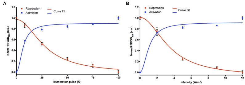
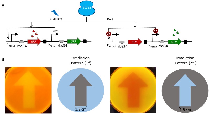
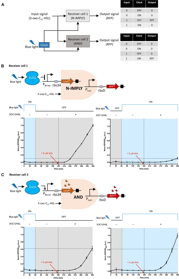
Light-regulated modules offer unprecedented new ways to control cellular behavior in precise spatial and temporal resolution. The availability of such tools may dramatically accelerate the progression of synthetic biology applications. Nonetheless, current optogenetic toolbox of prokaryotes has potential issues such as lack of rapid and switchable control, less portable, low dynamic expression and limited parts. To address these shortcomings, we have engineered a novel bidirectional promoter system for Escherichia coli that can be induced or repressed rapidly and reversibly using the blue light dependent DNA-binding protein EL222. We demonstrated that by modulating the dosage of light pulses or intensity we could control the level of gene expression precisely. We show that both light-inducible and repressible system can function in parallel with high spatial precision in a single cell and can be switched stably between ON- and OFF-states by repetitive pulses of blue light. In addition, the light-inducible and repressible expression kinetics were quantitatively analysed using a mathematical model. We further apply the system, for the first time, to optogenetically synchronize two receiver cells performing different logic behaviors over time using blue light as a molecular clock signal. Overall, our modular approach layers a transformative platform for next-generation light-controllable synthetic biology systems in prokaryotes.


Figures

Figure 1.
Figure 1.
Design and characterization of the blue light inducible and repressible gene expression system. (A) The blue light inducible promoter (PBLind-v1) consists of EL222 binding region fused to the luxI promoter. In the dark state (OFF), EL222 is unable to bind DNA and the transcription of the PBLind promoter is turned on only when exposed to blue light which triggers the EL222 transcription factor to bind to the upstream binding region and presumably recruits RNAP similar to LuxR-based transcriptional activators. (B) For the inducible promoter characterization experiments, cells were transformed with pBLind (reporter) and pEL222 (constitutively expressing EL222 protein) plasmids. As a control, we transformed cells only with pBLind plasmid. (C) The blue light repressible promoter (PBLrep-v1) consists of the EL222 binding region positioned between consensus −35 and −10 regions of RNAP binding site. The promoter is constitutive in dark and upon exposure to blue light, EL222 binds to the promoter region presumably causing steric hindrance to RNAP binding thereby repressing the RFP transcription. (D) For the repressible promoter characterization experiments, cells were transformed with pBLrep (reporter) and pEL222 (constitutively expressing EL222 protein) plasmids. As a control, we transformed cells only with pBLrep plasmid. For both the promoters, rbs34 is placed downstream of the promoter driving the RFP reporter as the output. For both (B) and (D), cells were kept in the dark or exposed to blue light (465 nm; 12 W/m2 intensity) for 6 h. Data are represented as mean ± S.D. (n = 3). Statistical significance of ****P < 0.0001 was calculated based on two-way ANOVA test.
Figure 2.
Figure 2.
Dose-response curves of blue light inducible and repressible promoters. (A) Varying blue light illumination pulse (%), 0% (OFF), 8.33% (5 s ON; 55 s OFF), 25% (15 s ON; 45 s OFF), 50% (30 s ON; 30 s OFF), 75% (45 s ON; 15 s OFF) and 100% (ON). (B) Varying blue light intensity levels (2, 5, 9 and 12 W/m2). The curve-fits are shown as solid lines to the promoter transfer function. All data (RFP/OD600) were normalized to the highest value obtained. The error bars indicate S.D. (n = 3).
Figure 3.
Figure 3.
Light-switchable activation and repression in a single cell. (A) Design and mode of action of the biregulatory promoter system (pBPar). The blue light inducible promoter (PBLind-v1) drives the expression of RFP reporter, which is placed in series with the blue light repressible promoter (PBLrep-v1) that controls the GFP expression. rbs34 was used downstream of both PBlind and PBLrep. In the presence of blue light (12 W/m2) transcription of the PBLind-v1 is activated expressing RFP, while the PBLrep-v1 is repressed. Conversely in the dark state, RFP expression is de-activated at the PBlind, while PBlrep constitutively expresses GFP. (B) Spatial control of gene expression. Cells transformed with pBPar and pEL222 plasmids, plated on an agar plate were illuminated with blue light through a photomask with two different spatial irradiation patterns for 18–20 h at 37°C. Images were taken under UV illumination. The width of the arrow is 1.8 cm.
Figure 4.
Figure 4.
Temporal control of gene expression by blue light based bidirectional promoter system in a single cell. Cells transformed with pBPar and pEL222 plasmids were exposed to blue light repeatably and reversibly in (A) OFF–ON–OFF–ON cycle for every 2 h over a period of 8 h and (E) OFF–ON cycle for every 3 h over a period of 6 h. The data (Fluo/OD600) were normalized to the highest value obtained. (B) and (F) are calculated synthesis rates (min−1) based on the normalized Fluo/OD600 values obtained. Error bars indicate S.D. (n = 3). The solid lines represent the model predicted temporal behavior. Model predicted (C) and (G) mRNA synthesis rates (min−1) and (D) and (H) mRNA abundance (μM). Grey areas represent dark state (‘OFF’) while the blue regions (illumination ‘ON’, 12 W/m2).
Figure 5.
Figure 5.
Optogenetic synchronization of two receiver cells performing a modular AND gate and a modular N-imply gate respectively. (A) Schematic representation of the optogenetic synchronization of both receiver cell[1], performing N-imply logic and receiver cell[2] performing AND gate logic using blue light as the clock signal. The input to both the receivers is AHL signal. The output signal is the RFP fluorescence. Receiver cell[1], produces the output signal only when the blue light (clock) is turned ‘OFF’ and AHL (input) is present (N-imply logic) while the receiver cell[2] output signal is produced only when both blue light (clock) is turned ‘ON’ and AHL (input) is present (AND logic). (B) Top, mode of action of N-imply logic in receiver cell[1]. pQSBLrep: LasRLVA is expressed under PBLrep-v1 promoter, while the output RFPLVA signal is under PLasI promoter. Bottom, characterization results of N-imply gate logic in receiver cell[1] co-transformed with pQSBLrep and pEL222 plasmids. (C) Top, mode of action of AND logic in receiver cell[2]. pQSBLind: LasRLVA is expressed under PBLind-v1 promoter, while the output RFPLVA signal is under PLasI promoter. Bottom, characterization results of AND gate logic in receiver cell[2] co-transformed with pQSBLrep and pEL222 plasmids. In both the receivers, LasR and RFP are destabilized version of the proteins with LVA degradation tag, denoted by *. rbs34 and rbsD are used downstream of PBLrep-v1/PBlind-v1 and PLasI respectively. The blue light clock signals used were, pulse[1]: 1 h ON, 5 h OFF; and pulse[2]: 1 h OFF, 5 h ON. The input signal AHL (5 μM) was added at the end of third hour of the experiment. The data (RFP/OD600) were normalized to the highest value obtained. Error bars indicate S.D. (n = 3).

References

    1. Lenz P., Søgaard-Andersen L. Temporal and spatial oscillations in bacteria. Nat. Rev. Micro. 2011;9:565–577. - PubMed
    1. Toettcher J.E., Voigt C.A., Weiner O.D., Lim W.A. The promise of optogenetics in cell biology: interrogating molecular circuits in space and time. Nat. Methods. 2011;8:35–38. - PMC - PubMed
    1. Bacchus W., Fussenegger M. The use of light for engineered control and reprogramming of cellular functions. Curr. Opin. Biotechnol. 2012;23:695–702. - PubMed
    1. Muller K., Weber W. Optogenetic tools for mammalian systems. Mol. Biosyst. 2013;9:596–608. - PubMed
    1. Lutz R., Bujard H. Independent and tight regulation of transcriptional units in Escherichia coli via the LacR/O, the TetR/O and AraC/I1-I2 regulatory elements. Nucleic Acids Res. 1997;25:1203–1210. - PMC - PubMed

MeSH terms

Substances