Review
. 2016 Aug 10;116(15):8693-749.
doi: 10.1021/acs.chemrev.6b00180. Epub 2016 Jun 29.

Hydrogenase Enzymes and Their Synthetic Models: The Role of Metal Hydrides

Affiliations
Review

Hydrogenase Enzymes and Their Synthetic Models: The Role of Metal Hydrides

David Schilter et al. Chem Rev. .

Abstract

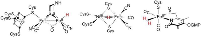
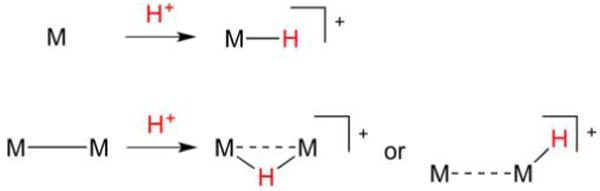
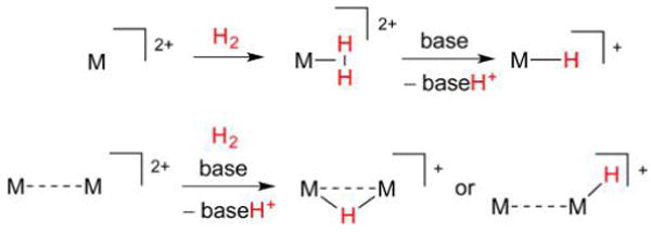
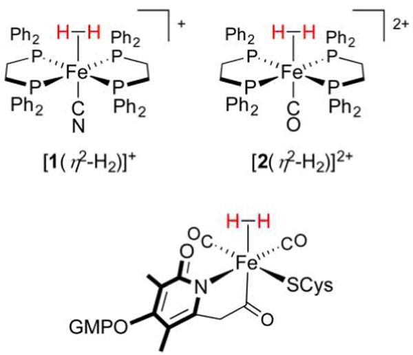
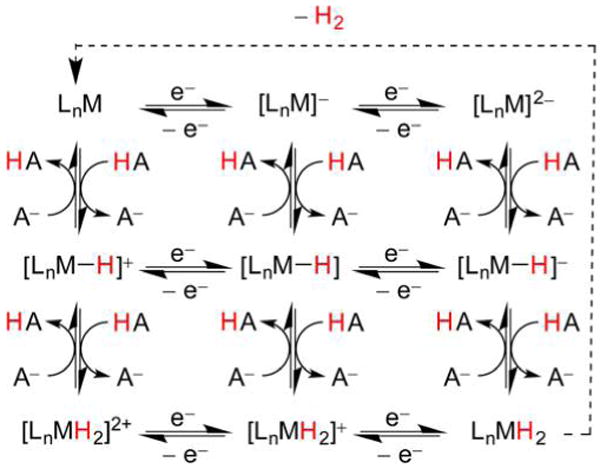
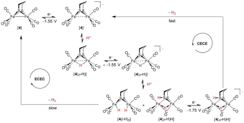
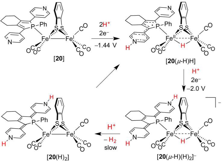
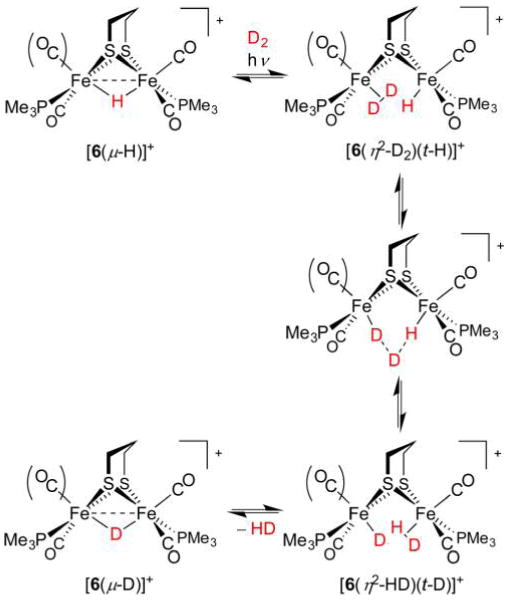
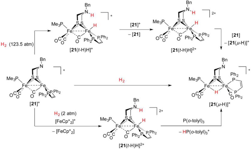
Hydrogenase enzymes efficiently process H2 and protons at organometallic FeFe, NiFe, or Fe active sites. Synthetic modeling of the many H2ase states has provided insight into H2ase structure and mechanism, as well as afforded catalysts for the H2 energy vector. Particularly important are hydride-bearing states, with synthetic hydride analogues now known for each hydrogenase class. These hydrides are typically prepared by protonation of low-valent cores. Examples of FeFe and NiFe hydrides derived from H2 have also been prepared. Such chemistry is more developed than mimicry of the redox-inactive monoFe enzyme, although functional models of the latter are now emerging. Advances in physical and theoretical characterization of H2ase enzymes and synthetic models have proven key to the study of hydrides in particular, and will guide modeling efforts toward more robust and active species optimized for practical applications.


Conflict of interest statement

Notes The authors declare no competing financial interest.

Figures

Figure 1
Figure 1
Schematic representations of the active sites in [FeFe]-H2ase (left), [NiFe]-H2ase (center), and [Fe]-H2ase (right). The presence of H and H2 ligands in [FeFe]-H2ase and [Fe]-H2ase, respectively, has yet to be confirmed.
Figure 2
Figure 2
Formation of mono- and dinuclear metal hydrides from low valent metals and H+. In the latter case, both bridging and terminal hydrido products are possible.
Figure 3
Figure 3
Interactions of low-valent metal sites with H2. The η2-H2 ligand can cleave through oxidative addition to afford dihydride complexes.
Figure 4
Figure 4
Interactions of high-valent metal sites with H2. The η2-H2 ligand in high-valent complexes can be acidic and is cleaved through deprotonation (heterolysis).
Figure 5
Figure 5
Fe(η2-H2) complexes in the laboratory (top left and right) and in Nature (bottom). Note the presence of five strong-field ligands that serve to support the low-spin Fe(II) electrophile.
Figure 6
Figure 6
Thermodynamic parameters for redox and acid–base processes for a metal complex M. Colored arrows denote transfer of an electron (blue), proton (red), H (black), and H (green). Adapted from ref . Copyright 2012 American Chemical Society.
Figure 7
Figure 7
X-ray structure of the [FeFe]-H2ase active site from Clostridium pasteurianum (PDB code 3C8Y). H atoms are omitted for clarity.
Figure 8
Figure 8
Reconstitution of apo-[FeFe]-H2ase from Chlamydomonas reinhardtii with [NC(CO)2Fe(adt)Fe(CO)2CN]2−.,
Figure 9
Figure 9
Catalytic cycle proposed for [FeFe]-H2ase (right) and electron counting for the [2Fe] unit in the three characterized states (left). The totals include Fe 3d electrons and bonding electron pairs from donor atoms. The substrate H atoms are in red for emphasis, although they cannot be distinguished from the amine H atoms.
Figure 10
Figure 10
Archetypal diiron thiolato hexacarbonyl [4] can be converted to its hydride [4(μ-H)]+ only with very strong acids. An alternative structural representation of [4(μ-H)]+, using full arrow and half-arrow notation, is provided below.
Figure 11
Figure 11
Catalytic cycle for electrocatalytic proton reduction mediated by [5(μ-H)].
Figure 12
Figure 12
The 1e reduction of a diamagnetic Fe(II)(μ-H)Fe(II) hydride affords a Fe(1.5)(μ-H)Fe(1.5) mixed-valent hydride.
Figure 13
Figure 13
Formation and isomerism of the mixed-valent hydride [7(μ-H)]. The μ-H ligand is unaffected by acid.
Figure 14
Figure 14
Preparation of the dihydride [8(μ-H)(t-H)] from sources of H+ and H.
Figure 15
Figure 15
Preparation of a dihydride through oxidative addition.
Figure 16
Figure 16
Preparation (top) and X-ray structures of [11(t-H)]+ and ([11(μ-H)]+ (bottom). Non-hydride H atoms are omitted for clarity.
Figure 17
Figure 17
Mechanisms for the isomerization of terminal to bridging hydride complexes.
Figure 18
Figure 18
Bridging secondary phosphido ligands do not represent sites for protonation, and only bridging hydrides are observed. In contrast, bridging thiolato ligands can be protonated, and serve as relays to afford terminal and bridging hydride complexes.
Figure 19
Figure 19
Rotated and unrotated isomers of [12], an Fe(I)Fe(I) model for Hred.
Figure 20
Figure 20
Protonation of [15] affords a thiol intermediate en route to a terminal hydride. Formation of the bridging hydride thermodynamic product is acid-catalyzed.
Figure 21
Figure 21
X-ray structures of [16(t-H)]+ and [17(t-H)H]2+. Nonionizable H atoms are omitted for clarity.,
Figure 22
Figure 22
Conformational dynamics accompanying the protonation of [17] (inversion of ammonium/amine centers not depicted).
Figure 23
Figure 23
Many proton and electron transfers that can be involved in the HER.
Figure 24
Figure 24
HER mechanism associated with [4].
Figure 25
Figure 25
HER mechanism associated with [18].
Figure 26
Figure 26
HER mechanism associated with [19].
Figure 27
Figure 27
Highly active HER catalyst [17] operates by different mechanisms that depend on acid strength.
Figure 28
Figure 28
HER catalytic cycle proposed for [20], which bears a redox-active phosphole ligand.
Figure 29
Figure 29
H/D scrambling mediated by [6(μ-H)]+. This complex may undergo either decarbonylation or μ-H → t-H isomerization such that a vacant site for D2 binding is accessible.
Figure 30
Figure 30
Stoichometric activation of H2 with [21]+ in the presence and absence of an external oxidant.
Figure 31
Figure 31
Catalytic cycle proposed for [23]-mediated H2 evolution and oxidation (clockwise and counterclockwise, respectively).,
Figure 32
Figure 32
Catalytic cycle proposed for H2 oxidation mediated by PNP complex [24(μ-H)]+.
Figure 33
Figure 33
X-ray structure of the [NiFe]-H2ase active site from Desulfovibrio vulgaris Miyazaki F (PDB code 4U9H). Nonionizable H atoms are omitted for clarity.
Figure 34
Figure 34
Catalytic cycles proposed for [NiFe]-H2ase. The electronic spin (S) and electron count of the NiFe cores (the sum of 3d electrons and electrons in metal–ligand bonds) are presented below each structure. A H2O ligand may be weakly bound to Ni in Ni-SIa.
Figure 35
Figure 35
Alternative mechanism for H2 activation by [NiFe]-H2ase, in which a proximal Arg residue (rather than a terminal Cys ligand) relays H+ to and from the active site.
Figure 36
Figure 36
Initial work on hydride-containing [NiFe]-H2ase models focused on the Fe subsite, whose Fe(CO)(CN)2H fragment is replicated by [25].
Figure 37
Figure 37
Synthesis of a Ni(I)Fe(I) thiolate [26], which can accept H+ either from acid or from H2 heterolysis to afford the hydride derivative [26(μ-H)]+.
Figure 38
Figure 38
X-ray structure of [26(μ-H)]+. Non-hydride H atoms are omitted for clarity.
Figure 39
Figure 39
Synthesis of phosphine-substituted Ni(pdt)(μ-H)Fe derivatives is best performed from hydrido tricarbonyl [26(μ-H)]+.
Figure 40
Figure 40
Synthesis of NiRu and NiW dithiolates and their respective hydrides.
Figure 41
Figure 41
X-ray structure of [36(μ-H)]+, one of the most active synthetic NiFe HER catalysts. Non-hydride H atoms are omitted for clarity.
Figure 42
Figure 42
HER catalytic cycle proposed for the present NiFe dthiolato hydrides.
Figure 43
Figure 43
Interconversion between thiol and hydride tautomers of [43H]+. There is evidence for both thiol and hydride moieties in the structure for Ni-R.
Figure 44
Figure 44
DFT-calculated structure for the putative species [44(μ-H)]+, whose Ni(III)(μ-H)Fe(II) core mimics that in Ni-C. Non-hydride H atoms omitted for clarity.
Figure 45
Figure 45
NiRu dithiolate [45(OH2)]2+ activates H2 to afford hydride [45(μ-H)(OH2)]+. Catalytic H2 oxidation, using Cu2+ as the electron acceptor, is proposed to involve a dihydride intermediate.
Figure 46
Figure 46
Activation of H2 by Ni(II)Fe(II) species [46(MeCN)]2+ affords hydride [46(μ-H)]+ (top). X-ray structure of [46(μ-H)]+ (bottom; non-hydride H atoms omitted for clarity).
Figure 47
Figure 47
Activation of H2 by in situ generated coordinatively unsaturated Ni(II)Fe(II) species affords hydride models for Ni-R (top). X-ray crystal structure of anion [47(μ-H)] (bottom; F and non-hydridic H atoms omitted for clarity).
Figure 48
Figure 48
H/D scrambling implies a Ni(η2-H2) intermediate (top). Such Ni(η2-H2) species can exhibit heterolysis with and without an external base (middle and bottom, respectively).
Figure 49
Figure 49
Examples of the [Ni(P2N2)2] family of electrocatalysts mediate both H2 oxidation (counterclockwise) and evolution (clockwise). This cycle is a simplification that neglects certain side pathways, and the order of the steps depends on the identities of substituents R and R′.
Figure 50
Figure 50
X-ray structure of the [Fe]-H2ase active site from Methanocaldococcus jannaschii (PDB code 3F4Z). H atoms are omitted for clarity.
Figure 51
Figure 51
Catalytic cycle proposed for [Fe]-H2ase.
Figure 52
Figure 52
Early models for CO-inhibited [Fe]-H2ase feature some of the donor groups present in the enzyme.,,
Figure 53
Figure 53
Acyl- and carbamoylpyridine models for CO-inhibited [Fe]-H2ase.
Figure 54
Figure 54
Pentacoordinate acylpyridine models [59] and [60] replicate inner Fe coordination sphere of the active [Fe]-H2ase states, yet do not bind H2.,
Figure 55
Figure 55
Preparation and reactivity of the Fe hydrido carbamoyl [61(H)H].
Figure 56
Figure 56
Polar hydrogenation of an aldehyde catalyzed by [63]+.
Figure 57
Figure 57
Complex [RuCp(CO)2] exhibits [Fe]-H2ase behavior, effecting H2 heterolysis and delivery of H to an imidazolium substrate resembling methenyl-H4MPT+.
Figure 58
Figure 58
Proton transport to and from the [2Fe] site in [FeFe]-H2ase (formal charges omitted). This involves H+ movement, denoted by red arrows, between Fed, the adt2− cofactor, and the protein (via Cys299), and likely requires pyramidal inversion of the amine.

References

    1. Immerdorf H. Contributions to the solution of the “nitrogen question”. Landwirtsch Jahrb. 1892;21:281.
    1. Tausz J, Donath H. On the oxidation of hydrogen and hydrocarbons by bacteria. Hoppe-Seyler’s Z Physiol Chem. 1930;190:141.
    1. Stephenson M, Stickland LH. XXVII. Hydrogenase: a bacterial enzyme activating molecular hydrogen. I. The properties of the enzyme. ochem J. 1931;25:205–214. - PMC - PubMed
    1. Elsden SR. Hydrogenase 1931–1981. Trends Biochem Sci. 1981;6:251–253.
    1. Stripp ST, Happe T. How algae produce hydrogen—news from the photosynthetic hydrogenase. Dalton Trans. 2009:9960–9969. - PubMed

LinkOut - more resources