. 2016 Jun 29:6:29024.
doi: 10.1038/srep29024.

Lactobacillus crispatus inhibits the infectivity of Chlamydia trachomatis elementary bodies, in vitro study

Affiliations

Lactobacillus crispatus inhibits the infectivity of Chlamydia trachomatis elementary bodies, in vitro study

Paola Nardini et al. Sci Rep. .

Abstract

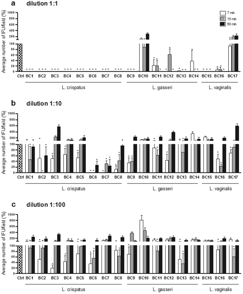
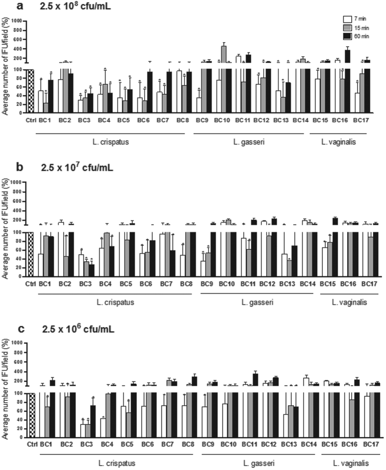
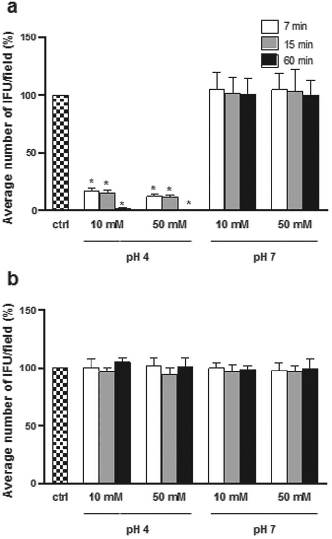
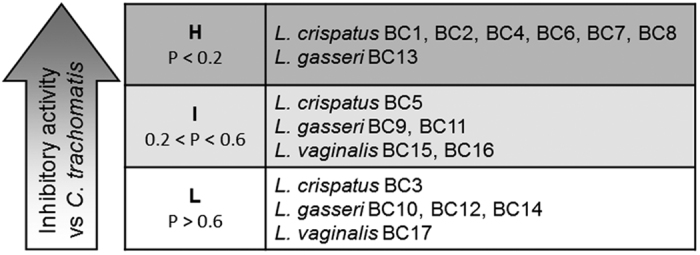
Lactobacillus species dominate the vaginal microbiota of healthy reproductive-age women and protect the genitourinary tract from the attack of several infectious agents. Chlamydia trachomatis, a leading cause of sexually transmitted disease worldwide, can induce severe sequelae, i.e. pelvic inflammatory disease, infertility and ectopic pregnancy. In the present study we investigated the interference of Lactobacillus crispatus, L. gasseri and L. vaginalis, known to be dominant species in the vaginal microbiome, with the infection process of C. trachomatis. Lactobacilli exerted a strong inhibitory effect on Chlamydia infectivity mainly through the action of secreted metabolites in a concentration/pH dependent mode. Short contact times were the most effective in the inhibition, suggesting a protective role of lactobacilli in the early steps of Chlamydia infection. The best anti-Chlamydia profile was shown by L. crispatus species. In order to delineate metabolic profiles related to anti-Chlamydia activity, Lactobacillus supernatants were analysed by (1)H-NMR. Production of lactate and acidification of the vaginal environment seemed to be crucial for the activity, in addition to the consumption of the carbonate source represented by glucose. The main conclusion of this study is that high concentrations of L. crispatus inhibit infectivity of C. trachomatis in vitro.


Figures

Figure 1
Figure 1. Effect of lactobacilli supernatants on C. trachomatis infectivity.
Experiments were performed with different dilutions of cell free supernatants: 1:1 (a), 1:10 (b) and 1:100 (c), and different time points: 7 minutes (white bars), 15 minutes (grey bars) and 60 minutes (black bars). C. trachomatis infectivity was evaluated as number of IFU/microscopic field. The results were expressed in percentage compared with control, taken as 100% (dotted bars). Bars represent median values, error bars represent median absolute deviations. Statistical significance was calculated vs control. *P < 0.05.
Figure 2
Figure 2. Effect of lactobacilli cell pellets on C. trachomatis infectivity.
Experiments were performed at different concentrations of cell pellets: 2.5 × 108 CFU/mL (a), 2.5 × 107 CFU/mL (b) and 2.5 × 106 CFU/mL (c), and different time points: 7 minutes (white bars), 15 minutes (grey bars) and 60 minutes (black bars). C. trachomatis infectivity was evaluated as number of IFU/microscopic field. The results were expressed in percentage compared with control, taken as 100% (dotted bars). Bars represent median values, error bars represent median absolute deviations. Statistical significance was calculated vs control. *P < 0.05.
Figure 3
Figure 3. Effect of lactic acid and hydrochloric acid on C. trachomatis infectivity.
Experiments were performed with lactic acid (a) and HCl (b). Infectivity was evaluated for different concentrations of lactic acid/HCl (10 mM and 50 mM), pH values (4 and 7), and time points [7 minutes (white bars), 15 minutes (grey bars) and 60minutes (black bars)]. C. trachomatis infectivity was evaluated as number of IFU/microscopic field. The results were expressed in percentage compared with control, taken as 100% (dotted bars). Bars represent median values, error bars represent median absolute deviations. Statistical significance was calculated vs control. *P < 0.05.
Figure 4
Figure 4. Ranking of lactobacilli in relation to anti-Chlamydia activity.
Lactobacillus strains were classified on the basis of the inhibitory activity of their cell free supernatants (CFS), expressed as the difference between CFS-treated EBs and untreated EBs, by means of the 1-tailed Wilcoxon signed rank P-values. Group H comprises lactobacilli strains with P-values below 0.2, group I comprises of lactobacilli with P-values ranging between 0.2 and 0.6; group L comprises lactobacilli with P-values over 0.6.
Figure 5
Figure 5. Correlation between metabolome of lactobacilli and inhibitory activity towards C. trachomatis.
(a) Score plot of Lactobacillus strains on PC1 and PC2 of a PCA model built on the total metabolites identified by 1H-NMR in cell free supernatants. H, I and L indicate the median values of lactobacilli grouped according to anti-Chlamydia activity. Strains without marks belong to group H; underlined strains belong to group I; strains within rectangles belong to group L. Expl. Var, explained variance. (b) Box plots representing the distribution of activity against Chlamydia in relation to the metabolome. Lines within the boxes indicate the median values of the samples groups corresponding to the different activity scores (H: high activity; I: intermediate activity; L: low activity). Each box represents the interquartile range (25–75th percentile). The bottom and top bars indicate the 10th and 90th percentiles, respectively.

Similar articles

Cited by

References

    1. Larsen B. & Monif G. R. Understanding the bacterial flora of the female genital tract. Clin. Infect. Dis. 32, e69–77 (2001). - PubMed
    1. Gupta K. et al. Inverse association of H2O2-producing lactobacilli and vaginal Escherichia coli colonization in women with recurrent urinary tract infections. J. Infect. Dis. 178, 446–450 (1998). - PubMed
    1. Pybus V. & Onderdonk A. B. Microbial interactions in the vaginal ecosystem, with emphasis on the pathogenesis of bacterial vaginosis. Microbes Infect. 1, 285–292 (1999). - PubMed
    1. Cherpes T. L., Meyn L. A., Krohn M. A., Lurie J. G. & Hillier S. L. Association between acquisition of herpes simplex virus type 2 in women and bacterial vaginosis. Clin. Infect. Dis. 37, 319–325 (2003). - PubMed
    1. Martin H. L. et al. Vaginal lactobacilli, microbial flora, and risk of human immunodeficiency virus type 1 and sexually transmitted disease acquisition. J. Infect. Dis. 180, 1863–1868 (1999). - PubMed

Publication types

MeSH terms