Review
. 2016 Sep;40(5):753-73.
doi: 10.1093/femsre/fuw013. Epub 2016 Jun 26.

Cyclic diguanylate signaling in Gram-positive bacteria

Affiliations
Review

Cyclic diguanylate signaling in Gram-positive bacteria

Erin B Purcell et al. FEMS Microbiol Rev. 2016 Sep.

Abstract

The nucleotide second messenger 3'-5' cyclic diguanylate monophosphate (c-di-GMP) is a central regulator of the transition between motile and non-motile lifestyles in bacteria, favoring sessility. Most research investigating the functions of c-di-GMP has focused on Gram-negative species, especially pathogens. Recent work in Gram-positive species has revealed that c-di-GMP plays similar roles in Gram-positives, though the precise targets and mechanisms of regulation may differ. The majority of bacterial life exists in a surface-associated state, with motility allowing bacteria to disseminate and colonize new environments. c-di-GMP signaling regulates flagellum biosynthesis and production of adherence factors and appears to be a primary mechanism by which bacteria sense and respond to surfaces. Ultimately, c-di-GMP influences the ability of a bacterium to alter its transcriptional program, physiology and behavior upon surface contact. This review discusses how bacteria are able to sense a surface via flagella and type IV pili, and the role of c-di-GMP in regulating the response to surfaces, with emphasis on studies of Gram-positive bacteria.

Keywords: Gram-positive; adherence; biofilm; cyclic diguanylate; motility; signaling.


Figures

Graphical Abstract Figure.
The majority of bacterial life exists in a surface-associated state, with motility allowing bacteria to disseminate and colonize new environments. Like in Gram-negative bacteria, c-di-GMP signaling in Gram-positives inversely controls the production of flagella and adherence factors and appears to be a major mechanism by which bacteria sense and respond to surfaces.
Figure 1.
Figure 1.
Growing interest in c-di-GMP signaling in Gram-positive bacteria. (A) The consensus of numerous studies indicates that c-di-GMP negatively regulates properties of free-living bacteria such as flagellum-mediated swimming, while promoting adherent phenotypes such as biofilm development. (B) Though c-di-GMP was first described in 1985 (Ross et al. 1985, 1987) and interest in c-di-GMP signaling exploded in 2004, c-di-GMP signaling in the context of Gram-positive bacteria went unreported before 2010 (den Hengst et al. 2010). Trends in publications including c-di-GMP were determined using the search engine at http://dan.corlan.net/medline-trend.html, with the search terms ‘c-di-GMP’ to identify the total number of publications. Search results for all years since 2009 were manually sorted to identify the subset of reports on Gram-positive bacteria. The numbers are likely to be underestimates given differences in terminology used for c-di-GMP in earlier studies (e.g. cyclic diguanylic acid, cyclic diguanylate, cyclic di-GMP).
Figure 2.
Figure 2.
Comparison of flagellum and TFP structures. (A) Diagram of the flagellum in Gram-positive bacteria, including key structural components of the basal body, hook and filament. Gram-positive bacteria have two basal body rings instead of four, one in the membrane and one in the peptidoglycan layer. The motor that drives flagellar rotation consists of stator proteins MotA and MotB, and rotor FliG, FliM and FliY (FliN). In the absence of ion flow through the motor, the stator and rotor complexes do not engage, and no flagellar rotation occurs. As ions flow though, dependent on adequate membrane potential, the motor and stator engage to generate the torque needed to propel the flagellum. In B. subtilis, the EpsE protein that contributes to biosynthesis of EPS production and biofilm formation also serves as a ‘clutch’, interacting with FliG to impede flagellar rotation. The PilZ domain protein YpfA/DgrA, a post-translational negative regulator of B. subtilis motility, interacts with MotA. (B) Diagram of the TFP of Gram-positive bacteria. The PilB ATPase powers the assembly of PilA pilin subunits into the base of the growing fiber; the PilT ATPase functions in disassembly of pilin subunits from the base, leading to retraction of the pilus as it shrinks. PilM, PilN and PilO comprise the membrane complex through which the pilus extends. In Gram-positive bacteria, the secretin that spans the outer membrane (PilQ in Gram-negatives) is absent, and the equivalent structure that allows the TFP to cross the peptidoglycan cell wall is unknown. (C–E) Comparisons of flagella (black arrowheads) and TFP (white arrowheads) on C. difficile by transmission electron microscopy Flagella measure ∼20 nm in diameter and can be two to three times as long as the cell (C, E). TFP are thinner and shorter, approximately 5–8 nm in diameter and up to several microns in length (D, E).
Figure 3.
Figure 3.
Control of flagellar gene expression by the Cd1 riboswitch in C. difficile. (A) Cd1 is a class I c-di-GMP riboswitch upstream of flgB, the first gene in a large (∼23 kb) flagellar operon in C. difficile. In the proposed model, in the absence of c-di-GMP, the Cd1 riboswitch structure allows synthesis of the full-length flagellar gene transcript, promoting biosynthesis of flagella and motility. In the presence of c-di-GMP (represented by the red circle), Cd1 folds into an alternate structure that includes a Rho-independent transcription terminator. This leads to destabilization of the RNAP-DNA complex, premature transcription termination, and reduced flagellar gene expression and motility. (B) The predicted structures of Cd1 with and without c-di-GMP (red circle) were determined based on alignment with the in vitro-characterized riboswitch Vc2 and using Mfold. The conserved stemloops are labeled (P2 and P3) (Sudarsan et al. ; Lee et al. 2010). Unlike Vc2, Cd1 is not predicted to form a P1 stem in the presence of c-di-GMP (Smith et al. 2009). The predicted contact residues for c-di-GMP, based on the sequence alignment with Vc2, are boxed. The predicted Rho-independent transcription terminator sequence is indicated with red text. The anti-terminator sequence complementary to a portion of the terminator (green text) is proposed to preclude formation of the transcription terminator, allowing transcription read-through.
Figure 4.
Figure 4.
Model of surface sensing via TFP in C. difficile. Intracellular c-di-GMP inhibits flagellar biosynthesis and stimulates TFP formation in C. difficile, both through riboswitch-dependent mechanisms controlling expression of the respective genes. Based on these data, we propose that flagella and TFP are produced under distinct spatiotemporal conditions in the large intestine, though there may be conditions under which both are produced. Peritrichous flagella may allow C. difficile (depicted as red bacilli) to swim through the viscous intestinal lumen and mucosa (a). TFP promote C. difficile biofilm formation, motility across solid surfaces and aggregation in vitro and may allow C. difficile to adhere to the intestinal epithelium (b) and/or other intestinal bacteria (c). Not shown, c-di-GMP also promotes the production of other putative adhesins, by directly promoting the transcription of the respective genes and/or inhibiting the production of the ZmpI protease that cleaves these adhesins (Cafardi et al. ; Soutourina et al. ; Hensbergen et al. ; Peltier et al. 2015). Decreases in c-di-GMP may thus restore flagellum biosynthesis and downregulate the putative adhesins, allowing C. difficile to detach and disseminate.
Figure 5.
Figure 5.
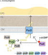
c-di-GMP promotes EPS production in L. monocytogenes. Listeria monocytogenes encodes three DGCs and three PDEs that control intracellular c-di-GMP levels (not shown). C-di-GMP positively regulates EPS production through PssE, an enzymatically inactive GGDEF domain protein that functions as a c-di-GMP receptor. The pssE gene is the fifth in a putative operon containing four genes predicted to be involved in EPS biosynthesis. PssC is a putative type 2 glycosyltransferase predicted to be involved in incorporation of the relevant sugars into the EPS. PssD has homology to BscB, the B subunit of the bacterial cellulose synthase. BscB does not catalyze cellulose synthesis, but are involved in export of the polymer. Thus, PssD may be involved in translocation of the as yet unknown EPS across the membrane. The remaining genes in the pssA-E locus, pssA and pssB, are predicted to encode a transmembrane protein and deacetylase, respectively. Their contributions to L. monocytogenes EPS biosynthesis have not been determined.

References

    1. Ababneh QO, Herman JK. RelA inhibits Bacillus subtilis motility and chaining. J Bacteriol. 2015;197:128–37. - PMC - PubMed
    1. Abee T, Kovacs AT, Kuipers OP, et al. Biofilm formation and dispersal in Gram-positive bacteria. Curr Opin Biotechnol. 2011;22:172–9. - PubMed
    1. Abel S, Bucher T, Nicollier M, et al. Bi-modal distribution of the second messenger c-di-GMP controls cell fate and asymmetry during the caulobacter cell cycle. PLoS Genet. 2013;9:e1003744. - PMC - PubMed
    1. Almblad H, Harrison JJ, Rybtke M, et al. The cyclic AMP-Vfr signaling pathway in Pseudomonas aeruginosa is inhibited by cyclic Di-GMP. J Bacteriol. 2015;197:2190–200. - PMC - PubMed
    1. Alteri CJ, Lindner JR, Reiss DJ, et al. The broadly conserved regulator PhoP links pathogen virulence and membrane potential in Escherichia coli. Mol Microbiol. 2011;82:145–63. - PMC - PubMed

Publication types

MeSH terms

Substances