. 2016 Aug 19;84(9):2616-26.
doi: 10.1128/IAI.00101-16. Print 2016 Sep.

Identification of Signaling Pathways by Which CD40 Stimulates Autophagy and Antimicrobial Activity against Toxoplasma gondii in Macrophages

Affiliations

Identification of Signaling Pathways by Which CD40 Stimulates Autophagy and Antimicrobial Activity against Toxoplasma gondii in Macrophages

Elizabeth Liu et al. Infect Immun. .

Abstract

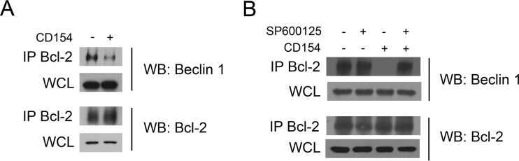
CD40 is an important stimulator of autophagy and autophagic killing of Toxoplasma gondii in host cells. In contrast to autophagy induced by nutrient deprivation or pattern recognition receptors, less is known about the effects of cell-mediated immunity on Beclin 1 and ULK1, key regulators of autophagy. Here we studied the molecular mechanisms by which CD40 stimulates autophagy in macrophages. CD40 ligation caused biphasic Jun N-terminal protein kinase (JNK) phosphorylation. The second phase of JNK phosphorylation was dependent on autocrine production of tumor necrosis factor alpha (TNF-α). TNF-α and JNK signaling were required for the CD40-induced increase in autophagy. JNK signaling downstream of CD40 caused Ser-87 phosphorylation of Bcl-2 and dissociation between Bcl-2 and Beclin 1, an event known to stimulate the autophagic function of Beclin 1. However, TNF-α alone was unable to stimulate autophagy. CD40 also stimulated autophagy via a pathway that included calcium/calmodulin-dependent kinase kinase β (CaMKKβ), AMP-activated protein kinase (AMPK), and ULK1. CD40 caused AMPK phosphorylation at its activating site, Thr-172. This effect was mediated by CaMKKβ and was not impaired by neutralization of TNF-α. CD40 triggered AMPK-dependent Ser-555 phosphorylation of ULK1. CaMKKβ, AMPK, and ULK1 were required for CD40-induced increase in autophagy. CD40-mediated autophagic killing of Toxoplasma gondii is known to require TNF-α. Knockdown of JNK, CaMKKβ, AMPK, or ULK1 prevented T. gondii killing in CD40-activated macrophages. The second phase of JNK phosphorylation-Bcl-2 phosphorylation-Bcl-2-Beclin 1 dissociation and AMPK phosphorylation-ULK1 phosphorylation occurred simultaneously at ∼4 h post-CD40 stimulation. Thus, CaMKKβ and TNF-α are upstream molecules by which CD40 acts on ULK1 and Beclin 1 to stimulate autophagy and killing of T. gondii.


Figures

FIG 1
FIG 1
JNK signaling is required for CD40-induced autophagy. (A) hmCD40 RAW 264.7 cells were incubated with or without CD154, followed by assessment of phospho-JNK and total JNK expression by immunoblotting. Densitometries of bands from CD154-treated cells were compared to those of bands from the corresponding control cells collected at the same time point. Densitometries for control bands for each time point were given a value of 1. Densitometry data represent means ± standard errors of the means of results from 3 experiments. (B) Mouse bone marrow-derived macrophages were incubated with CD154 and examined as described above. (C) hmCD40-RAW 264.7 cells transfected with tfLC3 were pretreated with SP600125 or vehicle for 1 h, followed by the addition of CD154. The expression of phospho-Thr183/Tyr185 JNK and total JNK was assessed by immunoblotting. The average numbers of autophagosomes (arrows) or autolysosomes (arrowheads) per cell were determined by fluorescence microscopy at 6 h. DMSO, dimethyl sulfoxide. (D) hmCD40-RAW 264.7 cells were pretreated with SP600125 or vehicle for 1 h, followed by the addition of CD154. Expression levels of p62/SQSTM1 and actin were assessed by immunoblotting at 24 h. (E) hmCD40-RAW 264.7 cells were transfected with control or JNK siRNA, followed by transfection with tfLC3. Total JNK and actin expression levels were assessed by immunoblotting. Average numbers of autophagosomes and autolysosomes per cell were determined as described above after 6 h of incubation with or without CD154. Results are shown as means ± standard errors of the means and are representative of data from 3 independent experiments. ***, P < 0.001.
FIG 2
FIG 2
TNF-α mediates the second phase of JNK phosphorylation after CD40 ligation and is required for CD40-induced autophagy. (A) hmCD40 RAW 264.7 cells were incubated with or without CD154 for 4 h. TNF-α production was examined by an ELISA. (B) hmCD40 RAW 264.7 cells transfected with tfLC3 were preincubated with an anti-TNF-α or isotype control MAb for 1 h, followed by the addition of CD154. The average numbers of autophagosomes and autolysosomes per cell were determined by fluorescence microscopy at 6 h. (C) hmCD40 RAW 264.7 cells were incubated with an anti-TNF-α or isotype control MAb, followed by the addition of CD154. Phospho-Thr183/Tyr185 JNK and total JNK were assessed by immunoblotting. (D) Bone marrow-derived macrophages from C57BL/6 (B6) and TNF-α−/− mice were incubated with CD154. Phospho-Thr183/Tyr185 JNK and total JNK levels were assessed by immunoblotting. Densitometries of bands from CD154-treated macrophages were compared to bands from their corresponding control macrophages. Densitometries for each control band were given a value of 1. Densitometry data represent means ± standard errors of the means of results from 3 experiments. Results shown are representative of data from 3 independent experiments. ***, P < 0.001.
FIG 3
FIG 3
CD40 ligation causes phosphorylation of Bcl-2 that is dependent on TNF-α and JNK. (A) hmCD40 RAW 264.7 cells were incubated with or without CD154. Phospho-Ser87 Bcl-2 and total Bcl-2 levels were assessed by immunoblotting. Densitometries of bands from CD154-treated cells were compared to those of bands from the corresponding control cells collected at the same time point. Densitometries for control bands for each time point were given a value of 1. (B and C) hmCD40 RAW 264.7 cells were incubated with anti-TNF-α versus an isotype control MAb (B) or SP600125 versus vehicle (C), followed by the addition of CD154. Phospho-Ser87 Bcl-2 and total Bcl-2 levels were assessed by immunoblotting at 4 h. Densitometry data represent means ± standard errors of the means of results from 3 experiments. Results are representative of data from 3 independent experiments.
FIG 4
FIG 4
CD40 ligation causes Bcl-2–Beclin 1 dissociation that is dependent on JNK signaling. (A) hmCD40-RAW 264.7 cells were incubated with or without CD154 for 4 h. Lysates were immunoprecipitated by incubation with an anti-Bcl-2 antibody and immunoblotted as indicated. Results are representative of data from 3 independent experiments. (B) hmCD40-RAW 264.7 cells were pretreated with SP600125 or vehicle for 1 h, followed by the addition of CD154 for 4 h. Lysates were subjected to immunoprecipitation (IP) and immunoblotting as described above. Results are representative of data from 3 independent experiments. WCL, whole-cell lysate; WB, Western blotting.
FIG 5
FIG 5
ULK1 is required for CD40-induced autophagy. (A) hmCD40 RAW 264.7 cells transfected with the tfLC3 plasmid were incubated with increasing concentrations of mouse recombinant TNF-α. The numbers of autophagosomes or autolysosomes were determined by fluorescence microscopy at 6 h. (B) hmCD40 RAW 264.7 cells were incubated with or without CD154, followed by assessment of phospho-Ser555 ULK1 and total ULK1 expression levels by immunoblotting. (C) hmCD40-RAW 264.7 cells were transfected with control or ULK1 siRNA, followed by transfection with tfLC3. ULK1 and actin expression levels were assessed by immunoblotting. The average numbers of autophagosomes and autolysosomes per cell were determined after 6 h of incubation with or without CD154. Expression levels of p62/SQSTM1 and actin were assessed by immunoblotting of cells transfected with control or ULK1 siRNA and incubated with or without CD154 for 24 h. Results are shown as means ± standard errors of the means and are representative of data from 3 independent experiments. ***, P < 0.001.
FIG 6
FIG 6
CaMKKβ-dependent AMPK signaling is required for CD40-induced autophagy. (A) hmCD40 RAW 264.7 cells were incubated with or without CD154, followed by assessment of phospho-Thr172 AMPK and total AMPK expression levels by immunoblotting. Densitometry data represent means ± standard errors of the means of results from 3 experiments. (B) hmCD40 RAW 264.7 cells were incubated with or without anti-TNF-α MAb, followed by stimulation with CD154. Total AMPK and phospho-Thr1174 AMPK levels were assessed by immunoblotting at 4 h. Densitometry data represent means ± standard errors of the means of results from 3 experiments. (C) hmCD40 RAW 264.7 cells were incubated with or without compound C (CC), followed by stimulation with human CD154. Expression levels of phospho-Ser555 ULK1 and total ULK1 were assessed by immunoblotting at 4 h. Densitometry data represent means ± standard errors of the means of results from 3 experiments. (D) hmCD40 RAW 264.7 cells were incubated with or without compound C, followed by stimulation with CD154. Expression levels of phospho-Ser 792 raptor and total raptor were assessed by immunoblotting at 4 h. (E) hmCD40-RAW 264.7 cells were transfected with the control or CaMKKβ. CaMKKβ and actin expression levels were assessed by immunoblotting. Total AMPK and phospho-Thr172 AMPK levels were assessed by immunoblotting 4 h after incubation with CD154. Densitometry data represent means ± standard errors of the means of results from 3 experiments. (F) hmCD40-RAW 264.7 cells transfected with tfLC3 were pretreated with STO-609, compound C, or vehicle for 1 h, followed by the addition of human CD154. The average numbers of autophagosomes or autolysosomes per cell were determined by fluorescence microscopy at 6 h. Expression levels of p62/SQSTM1 and actin were assessed by immunoblotting at 24 h. (G) hmCD40-RAW 264.7 cells were transfected with control, CaMKKβ, or AMPK1/2 siRNA, followed by transfection with tfLC3. Total AMPK and actin expression levels were assessed by immunoblotting. Numbers of autophagosomes and autolysosomes were determined as described above after 6 h of incubation with or without CD154. Results are shown as means ± standard errors of the means and are representative of data from 3 independent experiments. ***, P < 0.001.
FIG 7
FIG 7
JNK, CaMKKβ, AMPK, and ULK1 are required for CD40-induced killing of T. gondii. (A) hmCD40-RAW 264.7 cells were transfected with control siRNA or siRNA against JNK1/2, CaMKKβ, AMPK1/2, or ULK1, followed by incubation with or without CD154 and challenge with T. gondii. Percentages of infected macrophages and numbers of tachyzoites/100 macrophages were determined by light microscopy. (B) BMM from C57BL/6 mice were pretreated with STO-609 or vehicle for 1 h, followed by the addition of CD154 and challenge with T. gondii. Results are shown as means ± standard errors of the means and are representative of data from 2 to 3 independent experiments. **, P < 0.01.
FIG 8
FIG 8
Effects of CD40 signaling on ULK1 and Beclin 1. Shown is a schematic diagram illustrating the signaling pathways activated by CD40 that act on ULK1 and Beclin 1. Studies reported previously (21) revealed that CD40 ligation upregulates Beclin 1 protein levels through the reduction of p21 expression. Ser-555 ULK1 phosphorylation, Ser-87 Bcl-2 phosphorylation, and Beclin 1 upregulation appear to occur simultaneously. CD40 may activate additional mechanisms that act on ULK1 and Beclin 1 (see Discussion).

Similar articles

Cited by

References

    1. Mizushima N, Yoshimori T, Ohsumi Y. 2011. The role of Atg proteins in autophagosome formation. Annu Rev Cell Dev Biol 27:107–132. doi:10.1146/annurev-cellbio-092910-154005. - DOI - PubMed
    1. Itakura E, Mizushima N. 2010. Characterization of autophagosome formation site by a hierarchical analysis of mammalian Atg proteins. Autophagy 6:764–776. doi:10.4161/auto.6.6.12709. - DOI - PMC - PubMed
    1. Chan EY, Longatti A, McKnight NC, Tooze SA. 2009. Kinase-inactivated ULK proteins inhibit autophagy via their conserved C-terminal domains using an Atg13-independent mechanism. Mol Cell Biol 29:157–171. doi:10.1128/MCB.01082-08. - DOI - PMC - PubMed
    1. Russell RC, Tian Y, Yuan H, Park HW, Chang YY, Kim J, Kim H, Neufeld TP, Dillin A, Guan KL. 2013. ULK1 induces autophagy by phosphorylating Beclin-1 and activating VPS34 lipid kinase. Nat Cell Biol 15:741–750. doi:10.1038/ncb2757. - DOI - PMC - PubMed
    1. Egan DF, Shackelford DB, Mihaylova MM, Gelino S, Kohnz RA, Mair W, Vasquez DS, Joshi A, Gwinn DM, Taylor R, Asara JM, Fitzpatrick J, Dillin A, Viollet B, Kundu M, Hansen M, Shaw RJ. 2011. Phosphorylation of ULK1 (hATG1) by AMP-activated protein kinase connects energy sensing to mitophagy. Science 331:456–461. doi:10.1126/science.1196371. - DOI - PMC - PubMed

MeSH terms

LinkOut - more resources