. 2017 Jan 1;23(1):137-148.
doi: 10.1158/1078-0432.CCR-16-0870. Epub 2016 Jun 28.

Tumor-Derived CCL2 Mediates Resistance to Radiotherapy in Pancreatic Ductal Adenocarcinoma

Affiliations

Tumor-Derived CCL2 Mediates Resistance to Radiotherapy in Pancreatic Ductal Adenocarcinoma

Anusha Kalbasi et al. Clin Cancer Res. .

Abstract

Purpose: Local tumor growth is a major cause of morbidity and mortality in nearly 30% of patients with pancreatic ductal adenocarcinoma (PDAC). Radiotherapy is commonly used for local disease control in PDAC, but its efficacy is limited. We studied the impact of selectively intervening on radiotherapy-induced inflammation as an approach to overcome resistance to radiotherapy in PDAC.

Experimental design: PDAC cell lines derived from primary pancreatic tumors arising spontaneously in KrasLSL-G12D/+;Trp53LSL-R172H/+;Pdx-1 Cre mice were implanted into syngeneic mice and tumors were focally irradiated using the Small Animal Radiation Research Platform (SARRP). We determined the impact of depleting T cells and Ly6C+ monocytes as well as inhibiting the chemokine CCL2 on radiotherapy efficacy. Tumors were analyzed by flow cytometry and IHC to detect changes in leukocyte infiltration, tumor viability, and vascularity. Assays were performed on tumor tissues to detect cytokines and gene expression.

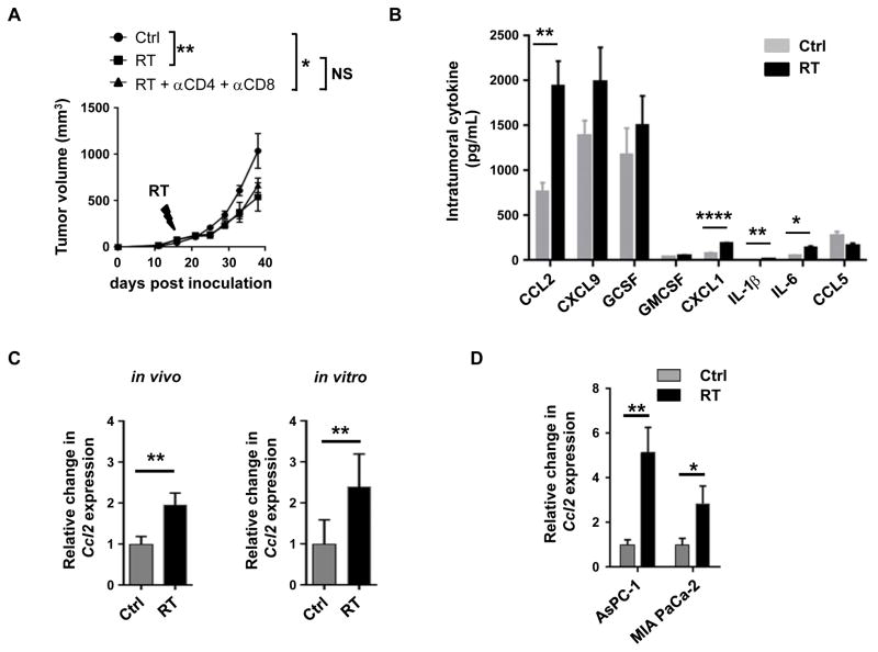
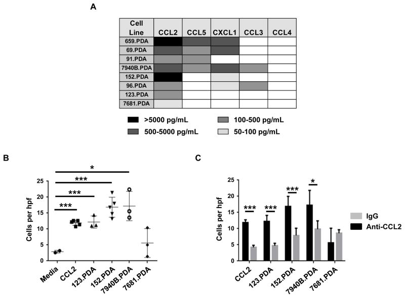
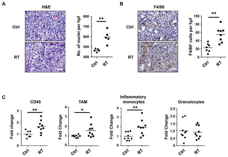
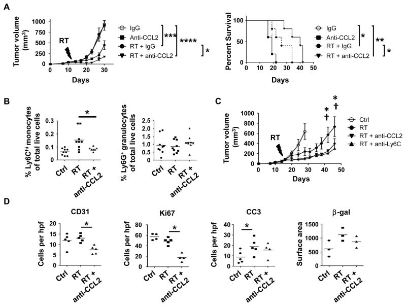
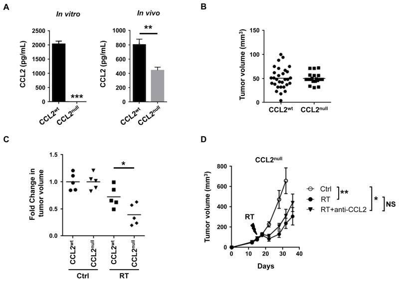
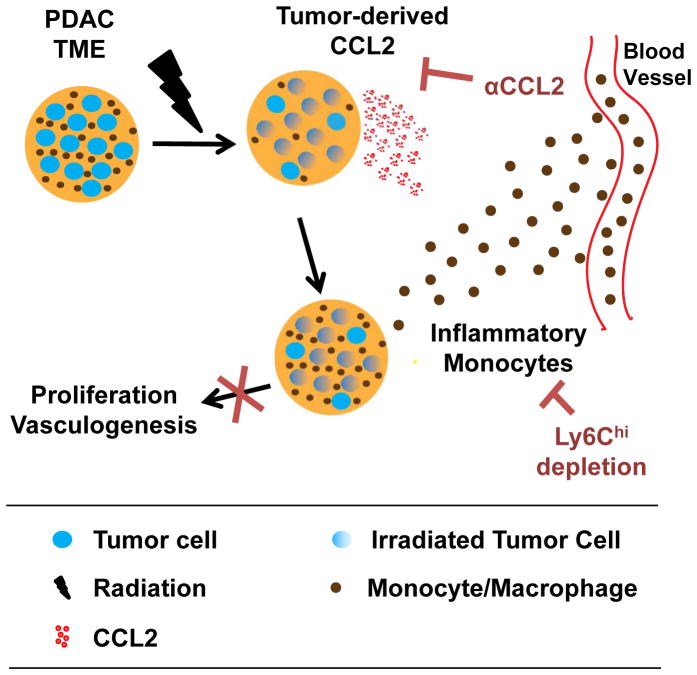
Results: Ablative radiotherapy alone had minimal impact on PDAC growth but led to a significant increase in CCL2 production by tumor cells and recruitment of Ly6C+CCR2+ monocytes. A neutralizing anti-CCL2 antibody selectively inhibited radiotherapy-dependent recruitment of monocytes/macrophages and delayed tumor growth but only in combination with radiotherapy (P < 0.001). This antitumor effect was associated with decreased tumor proliferation and vascularity. Genetic deletion of CCL2 in PDAC cells also improved radiotherapy efficacy.

Conclusions: PDAC responds to radiotherapy by producing CCL2, which recruits Ly6C+CCR2+ monocytes to support tumor proliferation and neovascularization after radiotherapy. Disrupting the CCL2-CCR2 axis in combination with radiotherapy holds promise for improving radiotherapy efficacy in PDAC. Clin Cancer Res; 23(1); 137-48. ©2016 AACR.


Conflict of interest statement

The authors have no competing financial interests.

Figures

Figure 1
Figure 1. RT-resistance in PDAC is associated with increased production of tumor-derived CCL2
(A) Tumor growth curve of a KPC-derived PDAC cell line (152.PDA) implanted subcutaneously and treated on day 16 after implantation with RT (20 Gy) or sham (Ctrl) in combination with CD4 and CD8 T cell depleting antibodies compared to isotype controls. Shown is mean ± s.e.m, n=5 mice per group. Tumor growth was compared using repeated measure two-way Anova (p<0.05) with Tukey multiple comparisons of means to evaluate differences between two groups. (B) Bar graph displaying intratumoral cytokine and chemokine levels (pg/mL) detected 24 hours after sham (Ctrl) or RT (20 Gy). (C) Fold change in Ccl2 expression detected within tumors in vivo and in vitro 3 days after RT (20 Gy) compared to sham (Ctrl). Ccl2 expression was normalized to β-actin. (D) Fold change in CCL2 expression detected in human PDAC cell lines AsPC-1 and MIA PaCa-2 at 3 days after RT compared to sham (Ctrl). For each treatment group, CCL2 expression was normalized to GAPDH. Significance testing was performed using unpaired two-tailed Student’s t test. *, p<0.05; **, p<0.01; ***, p<0.001; ****, p <0.0001.
Figure 2
Figure 2. PDAC-derived CCL2 induces myeloid cell migration
(A) Heat map displaying chemokine production by KPC-derived PDAC cell lines grown in vitro. (B) Migration of bone marrow-derived cells in response to tumor-derived soluble factors from indicated cell lines with comparison to CCL2 (10 ng/mL) and media alone. (C) Shown is the impact of anti-CCL2 neutralizing antibodies versus isotype control (IgG) on bone marrow-derived cell migration induced by tumor-derived soluble factors from indicated cell lines or CCL2 (10 ng/mL). For B and C, migrated cells were quantified at 40x magnification (hpf). Significance testing was performed using unpaired 2-tailed Student’s t test. *, p<0.05; **, p<0.01; ***, p<0.001; ****, p <0.0001.
Figure 3
Figure 3. RT enhances myeloid cell recruitment to the tumor microenvironment in PDAC
(A) Representative images of H&E staining of PDAC tumors (152.PDA) at 3 days after treatment with RT (20 Gy) versus sham. Nuclei are quantified as a measure of cellularity. (B) Representative images of F4/80 immunostaining of PDAC tumors (152.PDA) at 3 days after treatment with RT (20 Gy) versus sham (Ctrl). F4/80+ cells are quantified. For A and B, images are taken at 40X magnification (hpf) and scale bar represents 50 microns. (C) Leukocyte subsets within tumors at 3 days after RT (20 Gy) versus sham (Ctrl). Shown are the fold changes of total CD45+ cells, tumor-associated macrophages (TAM, CD45+CD11b+F480+Ly6Clo), inflammatory monocytes (CD45+CD11b+F4/80+Ly6Chi), and granulocytes (CD45+CD11b+F4/80negLy6G+) as a percentage of total live cells. Fold change is calculated relative to the average of the control group. Each data point represents a single mouse. Significance testing was performed using unpaired two-tailed Student’s t test. *, p<0.05; **, p<0.01.
Figure 4
Figure 4. CCL2 neutralization inhibits RT-induced recruitment of inflammatory monocytes to PDAC and enhances RT efficacy
Established PDAC tumors (152.PDA) implanted into syngeneic mice were treated with RT (14 Gy) or sham. Anti-CCL2 neutralizing antibodies, anti-Ly6C depleting antibodies or isotype control were administered on days -1, 0, 1, and 3 of RT. (A) Tumor growth curve and Kaplan Meier survival plot showing impact of CCL2 neutralizing antibodies on RT efficacy. Significance testing for tumor growth over time was performed using repeated measures two-way Anova (p<0.0001) with Tukey multiple comparisons of means as a post hoc test to evaluate differences between two groups. Survival significance was tested using log-rank analysis. (B) Shown are percentages of Ly6Chi inflammatory monocytes and Ly6G+ granulocytes of total live cells detected within tumors at 3 days after RT (20 Gy), versus sham (Ctrl), with or without anti-CCL2 neutralizing antibodies. Each data point represents a single mouse. (C) Tumor growth curve showing impact of anti-CCL2 and anti-Ly6C antibodies on RT efficacy. Significance testing was performed at each time point using Student’s t test with Holm-Šidák method to correct for multiple comparisons. *, p<0.05 for RT vs RT + anti-CCL2; †, p<0.05 for RT versus RT + anti-Ly6C. Data from panels A and C are representative of at least five and two independent experiments, respectively. (D) Quantification of immunohistochemical staining for vascularity (CD31), proliferation (Ki67), apoptosis (Cleaved Caspase 3), and senescence (β-galactosidase). Immunostains were quantified at 40x magnification (hpf). Each data point represents a single mouse. For panels B and D, significance testing was performed using unpaired 2-tailed Student’s t test. *, p<0.05; **, p<0.01; ***, p<0.001; ****, p <0.0001.
Figure 5
Figure 5. Tumor-derived CCL2 regulates RT efficacy in PDAC
CRISPR technology was used to knockout CCL2 in PDAC cells. (A) Shown is CCL2 (pg/mL) produced by wild-type PDAC cells (CCL2wt) and CCL2-knockout PDAC cells (CCL2null) in vitro and in vivo. (B) Tumor volume of CCL2wt and CCL2null PDAC cell lines at 12 days after implantation. Each data point represents a single mouse. (C) Fold change in tumor volume 4 days after ablative RT (14 Gy) in control (CCL2wt) and CCL2null clonal PDAC cell lines, relative to baseline. Data were normalized to untreated controls. (D) Tumor growth curve for CCL2null PDAC cells treated with RT (14 Gy), versus sham (Ctrl), with or without anti-CCL2 neutralizing antibodies. Shown is mean ± s.e.m; n=5 mice per group. Data are representative of 2 independent experiments. For panels A-C, significance testing was performed using unpaired two-tailed Student’s t test. For panel D, significance testing for tumor growth over time was determined using repeated measures two-way Anova (p<0.0001) with Tukey multiple comparisons of means as a post hoc test to evaluate differences between two groups. *, p<0.05; **, p <0.01, ***, p<0.001.
Figure 6
Figure 6. Conceptual model describing a role for monocytes and CCL2 in PDAC resistance to RT
The PDAC tumor microenvironment (TME) consists of tumor cells (blue), leukocytes (brown) including macrophages and their precursor monocytes, and other non-malignant stromal cells including fibroblasts and endothelial cells (not shown). Ablative RT results in cell death in a fraction of tumor cells and leukocytes within the TME. PDAC cells respond to RT by increasing their production of chemokines, including CCL2, which acts to recruit CCR2+ inflammatory monocytes to the TME. Tumor-infiltrating monocytes/macrophages then support tumor proliferation and neovascularization and therein establish PDAC resistance to RT. Neutralization of CCL2 or depletion of Ly6Chi inflammatory monocytes blocks RT-induced monocyte/macrophage recruitment to tumors and their subsequent pro-tumor effects leading to enhanced RT efficacy.

References

    1. Ryan D, Hong T, Bardeesy N. Pancreatic adenocarcinoma. N Engl J Med. 2014;371:1039–49. - PubMed
    1. American Cancer Society. Cancer Facts & Figures 2016. Atlanta: Am. Cancer Soc; 2016.
    1. Iacobuzio-Donahue Ca, Fu B, Yachida S, Luo M, Abe H, Henderson CM, et al. DPC4 gene status of the primary carcinoma correlates with patterns of failure in patients with pancreatic cancer. J Clin Oncol. 2009;27:1806–13. - PMC - PubMed
    1. Wyse JM, Carone M, Paquin SC, Usatii M, Sahai AV. Randomized, double-blind, controlled trial of early endoscopic ultrasound-guided celiac plexus neurolysis to prevent pain progression in patients with newly diagnosed, painful, inoperable pancreatic cancer. J Clin Oncol. 2011;29:3541–6. - PubMed
    1. Moss AC, Morris E, Mac Mathuna P. Palliative biliary stents for obstructing pancreatic carcinoma. Cochrane Database Syst Rev. 2006:CD004200. - PubMed

MeSH terms