. 2016 Jul 12;113(28):7894-9.
doi: 10.1073/pnas.1605015113. Epub 2016 Jun 27.

Bacterial lipids activate, synergize, and inhibit a developmental switch in choanoflagellates

Affiliations

Bacterial lipids activate, synergize, and inhibit a developmental switch in choanoflagellates

Arielle Woznica et al. Proc Natl Acad Sci U S A. .

Abstract

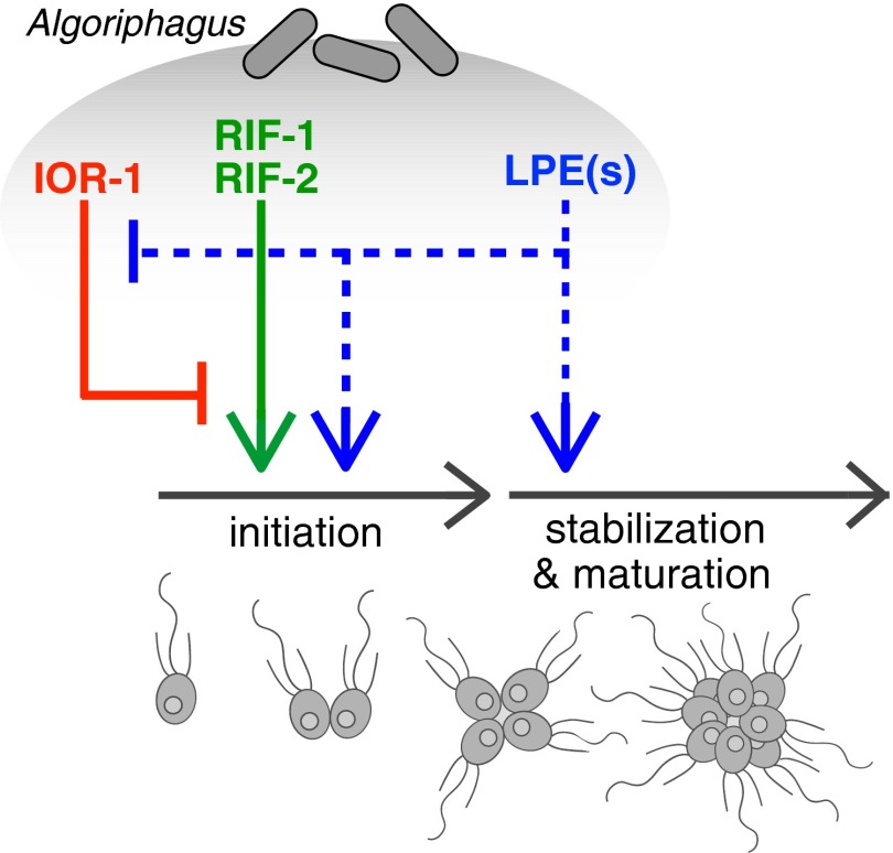
In choanoflagellates, the closest living relatives of animals, multicellular rosette development is regulated by environmental bacteria. The simplicity of this evolutionarily relevant interaction provides an opportunity to identify the molecules and regulatory logic underpinning bacterial regulation of development. We find that the rosette-inducing bacterium Algoriphagus machipongonensis produces three structurally divergent classes of bioactive lipids that, together, activate, enhance, and inhibit rosette development in the choanoflagellate Salpingoeca rosetta. One class of molecules, the lysophosphatidylethanolamines (LPEs), elicits no response on its own but synergizes with activating sulfonolipid rosette-inducing factors (RIFs) to recapitulate the full bioactivity of live Algoriphagus. LPEs, although ubiquitous in bacteria and eukaryotes, have not previously been implicated in the regulation of a host-microbe interaction. This study reveals that multiple bacterially produced lipids converge to activate, enhance, and inhibit multicellular development in a choanoflagellate.

Keywords: bacteria; choanoflagellates; host–microbe; multicellularity; sulfonolipid.


Conflict of interest statement

The authors declare no conflict of interest.

Figures

Fig. 1.
Fig. 1.
Stages of rosette development in S. rosetta. During rosette development, a single founding cell undergoes serial rounds of cell division, resulting in a structurally integrated rosette. Importantly, rosette development does not involve cell aggregation. Shown are a single cell (A), a pair of cells (B), a 4-cell rosette (C), an 8-cell rosette (D) and a 16-cell rosette (E).
Fig. 2.
Fig. 2.
Maximal rosette development requires lipid cofactor interactions. (A) When treated with media that lack necessary bacterial signals (Media Control), S. rosetta does not produce rosettes. In contrast, when treated with live Algoriphagus, Algoriphagus-conditioned media, OMVs from Algoriphagus, or bulk lipids extracted from Algoriphagus, rosettes develop at maximal (∼90% cells in rosettes) or near-maximal levels. (B) A heat map depicts the rosette-inducing activity of Algoriphagus lipid fractions used to treat SrEpac, either in isolation or in combination, at a final lipid concentration of 2 μg/mL Sulfonolipid-enriched fraction 11 was the only fraction sufficient to induce rosette development when tested alone (30% of cells in rosettes). Tests of each of the lipid fractions in combination revealed previously unidentified inhibitory and enhancing activity. Fractions 4 and 5 decreased rosette development (to 12% and 8%, respectively) in fraction 11-treated cells, whereas fraction 7 increased rosette development to 65%. (C) The RIF mix (solid square) and purified RIF-2 (solid circle) induced rosette development at micromolar concentrations. (Inset) RIF-1 (open circle) is active at femtomolar to nanomolar concentrations, but induces 10-fold lower levels of rosette development than RIF-2. The long gray box in the main graph indicates the range of concentrations at which RIF-1 is active and the range of its rosette-inducing activity. Rosette development was quantified 24 h after induction. Minor ticks on x axis are log-spaced.
Fig. 3.
Fig. 3.
Structural similarities and differences among RIFs, an inactive sulfonolipid, and the inhibitory capnine IOR-1. (A) The 3D structure of RIF-1 (43), compared with the proposed molecular structure of RIF-2, and (B) the structure of an inactive Algoriphagus sulfonolipid, Sulfobacin F (43). Shared features of Algoriphagus sulfonolipids include a fatty acid chain (shown in gray) and a capnoid base (shown in black). Distinguishing features between RIF-1 and RIF-2 (highlighted in red) include a double bond at position 4 and a hydroxyl group at position 6. The tight structure–activity relationships of RIF-1 and RIF-2 suggest a restricted set of interactions between these molecules and a binding target. No features are shared by RIF-1 and RIF-2 to the exclusion of Sulfobacin F. (C) The IOR-1 capnine antagonizes the rosette-inducing activity of RIFs. Like the capnoid base of RIF sulfonolipids, IOR-1 is composed of a sulfonic acid head group and a branched chain containing two -OH groups. Furthermore, the carbon chain length and branching pattern of IOR-1 matches the capnoid base in RIF-1 and -2. The similarities between IOR-1 and the RIFs raise the possibility that IOR-1 competitively inhibits RIF binding to a target receptor.
Fig. 4.
Fig. 4.
LPEs synergize with RIFs to enhance rosette development. (A) The structures of LPE 451 and LPE 465 as determined by NMR and tandem mass spectrometry. LPE 451 and LPE 465 differ by only one methyl group (highlighted in red). (B) The addition of 2 μM LPE mix increases the maximal percentage of cells in rosettes in RIF-2–treated SrEpac from 10.5% (solid circle) to 52% (open circle) and the maximal inducing activity of the RIF mix from 23.5% (solid square) to 82% (open square) of cells in rosettes. Minor ticks on x axis are log-spaced.
Fig. 5.
Fig. 5.
LPEs promote proper rosette development and maturation. (A) Frequency distribution of rosette size in SrEpac incubated with OMVs, RIF-2, and RIF-2 + LPE mix after exposure to shear stress by pipetting. Rosettes induced by RIF-2 alone contained fewer cells on average and reached a smaller maximal size than rosettes induced with OMVs isolated from Algoriphagus-conditioned media. The addition of the LPE mix to RIF-2 increased the median rosette size and frequency distribution to levels that recapitulated induction by OMVs. Rosette size was assessed 22 h after induction (n = 139 for each condition). Violin box plots show the median cell number (white circle), 75% quartile (thick line), and range excluding outliers (thin line). Surrounding the box plot is a kernel density trace, plotted symmetrically to show rosette size frequency distribution. P values (unpaired t tests) were calculated using GraphPad Prism v6 for Mac, GraphPad Software. (BE) Rosette morphology, cell packing, and localization of Rosetteless protein (a marker of rosette development) in rosettes induced by (B) OMVs, (C and D) RIF-2 alone, and (E) RIF-2 + LPEs. (B) Cells in OMV-induced rosettes express Rosetteless and are tightly packed. Anti-tubulin antibodies (white) highlight the cell body, and anti-Rosetteless antibodies (magenta) stain the Rosetteless protein in the center of rosettes (32). (C) Four-celled rosettes induced by RIF-2 are tightly packed, whereas larger rosettes induced by RIF-2 alone (D) appear disorganized with cells spaced farther apart. (E) Rosettes induced by the RIF-2 + LPE mix are large and closely packed and phenocopy rosettes induced by OMVs. All rosettes were fixed 22 h after treatment.
Fig. 6.
Fig. 6.
Multiple bacterial inputs regulate rosette development in S. rosetta. Algoriphagus produces three chemically distinct classes of lipids—sulfonolipids, LPEs, and a capnine—that interact to alternately induce, enhance, or inhibit rosette development in S. rosetta. The sulfonolipids RIF-1 and RIF-2 are sufficient to initiate rosette development in S. rosetta, although rosettes induced by RIFs alone are restricted in size, potentially because of their sensitivity to shear. Complete rosette maturation requires the synergistic activities of RIFs and LPEs. Although LPEs have no detectable activity on their own, they enhance RIF activity and facilitate the growth of larger and more structurally stable rosettes, perhaps by regulating downstream pathways important for rosette maturation. Although the molecular mechanisms by which LPEs regulate rosette development are unknown (indicated by dashed lines), multiple lines of evidence suggest that LPEs act both to promote the initiation of rosette development and, separately, to promote the subsequent maturation of rosettes. Algoriphagus also produces the inhibitory molecule IOR-1, which inhibits the rosette-inducing activity of RIFs (44). Importantly, when S. rosetta is exposed simultaneously to RIFs and the synergistic LPEs, mature rosettes develop even in the presence of IOR-1.

References

    1. Carroll SB. Chance and necessity: The evolution of morphological complexity and diversity. Nature. 2001;409(6823):1102–1109. - PubMed
    1. Tyler S. Epithelium: The primary building block for metazoan complexity. Integr Comp Biol. 2003;43(1):55–63. - PubMed
    1. King N. The unicellular ancestry of animal development. Dev Cell. 2004;7(3):313–325. - PubMed
    1. Stanley SM. An ecological theory for the sudden origin of multicellular life in the late precambrian. Proc Natl Acad Sci USA. 1973;70(5):1486–1489. - PMC - PubMed
    1. McFall-Ngai M. The development of cooperative associations between animals and bacteria: Establishing détente among domains. Am Zool. 1998;38(4):592–608.

MeSH terms