. 2016 Jul 12;113(28):7804-9.
doi: 10.1073/pnas.1606751113. Epub 2016 Jun 27.

Biomechanics of red blood cells in human spleen and consequences for physiology and disease

Affiliations

Biomechanics of red blood cells in human spleen and consequences for physiology and disease

Igor V Pivkin et al. Proc Natl Acad Sci U S A. .

Erratum in

Abstract

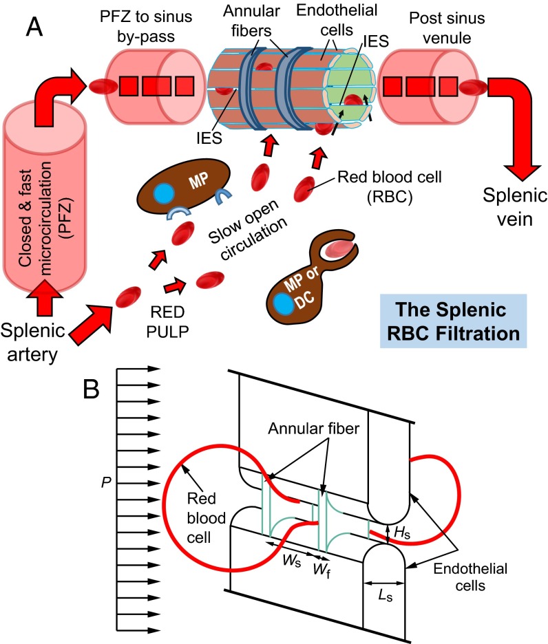
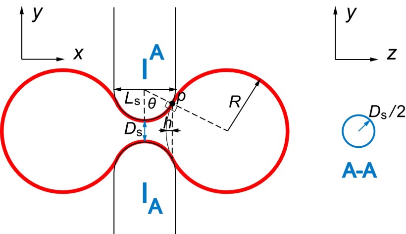
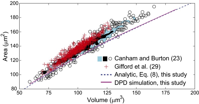
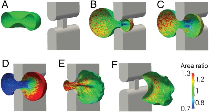
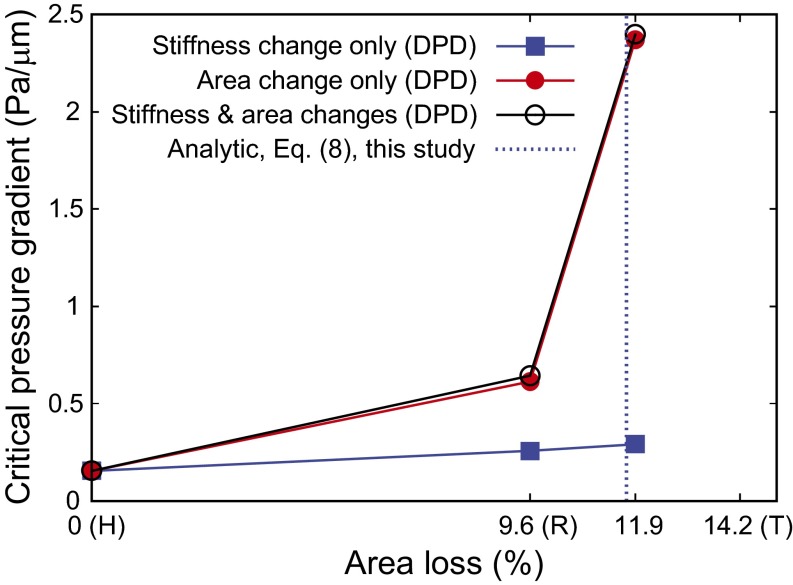
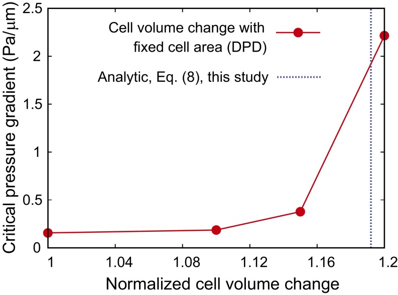
Red blood cells (RBCs) can be cleared from circulation when alterations in their size, shape, and deformability are detected. This function is modulated by the spleen-specific structure of the interendothelial slit (IES). Here, we present a unique physiological framework for development of prognostic markers in RBC diseases by quantifying biophysical limits for RBCs to pass through the IES, using computational simulations based on dissipative particle dynamics. The results show that the spleen selects RBCs for continued circulation based on their geometry, consistent with prior in vivo observations. A companion analysis provides critical bounds relating surface area and volume for healthy RBCs beyond which the RBCs fail the "physical fitness test" to pass through the IES, supporting independent experiments. Our results suggest that the spleen plays an important role in determining distributions of size and shape of healthy RBCs. Because mechanical retention of infected RBC impacts malaria pathogenesis, we studied key biophysical parameters for RBCs infected with Plasmodium falciparum as they cross the IES. In agreement with experimental results, surface area loss of an infected RBC is found to be a more important determinant of splenic retention than its membrane stiffness. The simulations provide insights into the effects of pressure gradient across the IES on RBC retention. By providing quantitative biophysical limits for RBCs to pass through the IES, the narrowest circulatory bottleneck in the spleen, our results offer a broad approach for developing quantitative markers for diseases such as hereditary spherocytosis, thalassemia, and malaria.

Keywords: erythrocytes; malaria; microcirculation; spherocytosis; spleen clearance.


Conflict of interest statement

The authors declare no conflict of interest.

Figures

Fig. 1.
Fig. 1.
(A) Illustration of the splenic RBC filtration function (adapted from ref. 18). Blood flow is from lower left to upper right and may follow two parallel paths. The closed and fast circulation goes from the splenic artery to central arterioles and their branches and then through the perifollicular zone (PFZ), PFZ-to-sinus bypasses, sinus lumens, and post-sinus venules that converge in the splenic vein, which consists of 80–90% of the splenic blood flow. The open and slow circulation through the red pulp involves (i) a microcirculatory structure without endothelial cells where cord macrophages (MPs) and reticular cells screen the slowly traversing blood cells and (ii) narrow and short IES in the sinus wall that RBCs must cross to go back to the general circulation. Less deformable RBCs retained mechanically by the IES and abnormal RBCs identified can be removed through phagocytosis by macrophages (MPs) or dendritic cells (DCs). (B) Computational model geometry setup of an RBC passing through an IES with a height of Hs, width of Ws, and length of Ls, which is divided by annular fibers with a fiber width of Wf. This process is driven by the hydrodynamic pressure gradient P.
Fig. 2.
Fig. 2.
Schematic illustration of the limiting geometry considered in the theoretical framework. The red solid line represents the RBC. At point p, the RBC surface is tangential to the surfaces of the endothelial cells. The cross-section A-A is shown on the right, where the solid line circle represents the slit cross-section. We approximate the real rectangular slit with the circular slit with the same cross-sectional area.
Fig. 3.
Fig. 3.
Predicted relationship between healthy RBC cell volume versus surface area for a pressure gradient of 1 Pa/µm. Solid curve, prediction by DPD simulations; dashed curve, prediction by the analytical theory, Eq. 8. The scatter circle points representing individual cells and shaded areas representing two different densities (blue and black regions representing less than and greater than three cells per 1 µm5, respectively) denote the experimental measurements of Canham and Burton (23). The red data points are from the experiments of Gifford et al. (29). Healthy RBCs with volumes and areas to the left of these curves would cross the splenic slits, whereas of RBCs located to the right of the curves would be retained at the IES.
Fig. 4.
Fig. 4.
DPD simulation of a sequence of six steps (AF) as an iRBC parasitized by P. falciparum passes through a slit in the human spleen at a constant pressure gradient of 0.64 Pa/µm. During this large deformation process, the iRBC is allowed to have a change up to 7% in total surface area from its undeformed value of 122 µm2. Only one-half of the RBC is shown for clarity and visualization. The color contours show local values of the ratio of the deformed to undeformed surface area of the RBC membrane. Here, area expansion occurs for values >1.0, whereas compression occurs for values <1.0.
Fig. 5.
Fig. 5.
The critical minimum pressure gradient for P. falciparum-infected RBCs in different stages to pass through the IES, as predicted by DPD. The blue dashed line is the limiting condition predicted by theory, Eq. 8, for critical area loss at fixed cell volume. Beyond this limiting geometry, an infinitely high pressure gradient is required for the RBCs to clear the splenic slit. The surface area loss for healthy (H), ring (R), and trophozoite (T) RBCs are taken to be 0%, 9.6%, and 14.2%, respectively, from the experiments of Safeukui et al. (13).
Fig. 6.
Fig. 6.
Dependence of the critical pressure gradient on the cell volume for fixed cell surface area. Blue dashed lines are the limiting geometries predicted by the theory Eq. 8.

References

    1. Mebius RE, Kraal G. Structure and function of the spleen. Nat Rev Immunol. 2005;5(8):606–616. - PubMed
    1. Petroianu A. The Spleen. Bentham Science Publishers; Hilversum, The Netherlands: 2011.
    1. Jandl JH, Greenberg MS, Yonemoto RH, Castle WB. Clinical determination of the sites of red cell sequestration in hemolytic anemias. J Clin Invest. 1956;35(8):842–867. - PMC - PubMed
    1. Waugh RE, et al. Rheologic properties of senescent erythrocytes: Loss of surface area and volume with red blood cell age. Blood. 1992;79(5):1351–1358. - PubMed
    1. Suresh S. Mechanical response of human red blood cells in health and disease: Some structure-property-function relationships. J Mater Res. 2006;21(8):1871–1877.

Publication types

LinkOut - more resources