. 2016 Jul;28(7):1563-80.
doi: 10.1105/tpc.16.00121. Epub 2016 Jun 27.

The Slicer Activity of ARGONAUTE1 Is Required Specifically for the Phasing, Not Production, of Trans-Acting Short Interfering RNAs in Arabidopsis

Affiliations

The Slicer Activity of ARGONAUTE1 Is Required Specifically for the Phasing, Not Production, of Trans-Acting Short Interfering RNAs in Arabidopsis

Laura Arribas-Hernández et al. Plant Cell. 2016 Jul.

Abstract

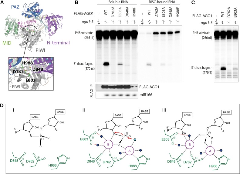
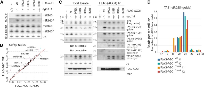
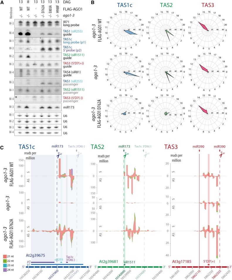
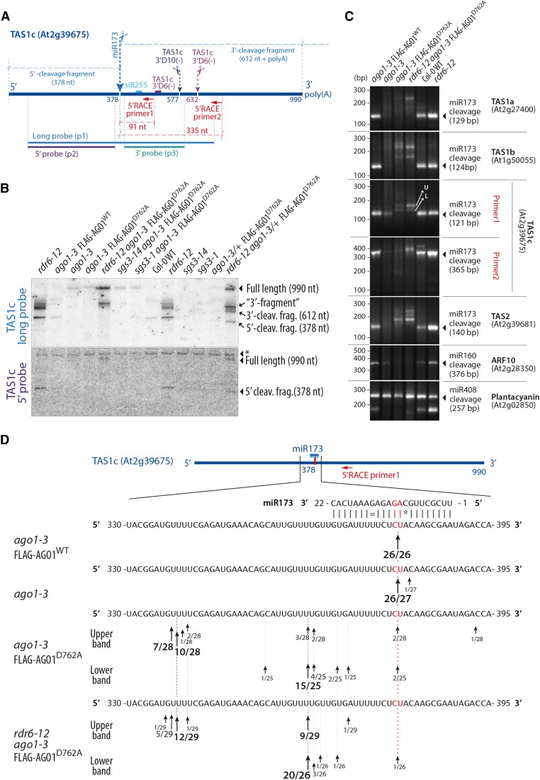
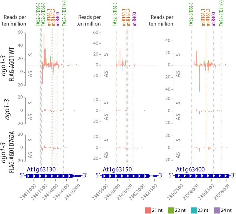
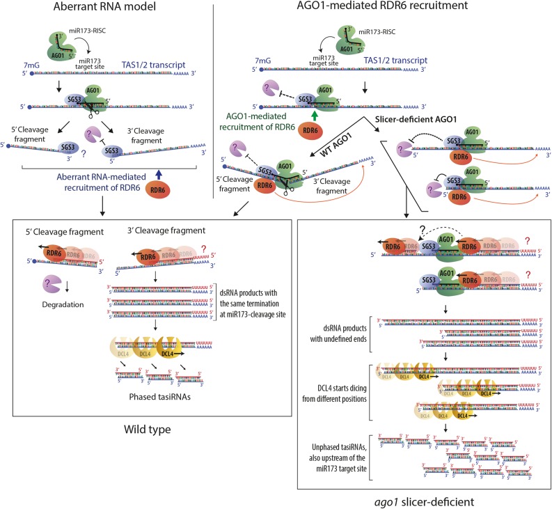
ARGONAUTE1 (AGO1) mediates posttranscriptional silencing by microRNAs (miRNAs) and short interfering RNAS (siRNAs). AGO1-catalyzed RNA cleavage (slicing) represses miRNA targets, but current models also highlight the roles of slicing in formation of siRNAs and siRNA-AGO1 complexes. miRNA-guided slicing is required for biogenesis of phased, trans-acting siRNAs (tasiRNAs), whose cleaved precursor fragments are converted to double-stranded RNA by RNA-dependent RNA polymerase 6 (RDR6). In addition, unwinding of duplex siRNA bound to AGO1 requires passenger strand cleavage in vitro. In this study, we analyze how mutation of four metal ion-coordinating residues of Arabidopsis thaliana AGO1 affects slicer activity in vitro and siRNA function in vivo. We show that while all four residues are required for slicer activity, they do not contribute equally to catalysis. Moreover, passenger strand cleavage is required for assembly of active AGO1-siRNA complexes in vivo, and many AGO1-bound siRNAs are trimmed in the absence of slicer activity. Remarkably, seedlings defective in AGO1 slicer activity produce abundant siRNAs from tasiRNA loci in vivo. These siRNAs depend on RDR6 and SUPPRESSOR OF GENE SILENCING3, but unlike wild-type tasiRNAs, they are unphased. These results demonstrate that slicing is solely required for phase definition of tasiRNAs, and they strongly support recruitment of RDR6 by AGO1 rather than by cleavage fragments.


Figures

Figure 1.
Figure 1.
Different Catalytic Roles of the Four Mg2+-Coordinating Active-Site Residues. (A) Structural model of AGO1 based on the crystal structure of human Ago2 (PDB ID 4EI3). The locations of the four residues involved in coordination of Mg2+-ions are indicated. (B) Slicer activity of immuno-affinity-purified FLAG-AGO1 from Arabidopsis inflorescences of stable transgenic lines expressing wild-type and slicer-deficient FLAG-AGO1. A 266-nucleotide 32P cap-labeled PHB single-stranded RNA bearing the endogenous miR166 target site was used as substrate. FLAG-AGO1 was immobilized on M2-conjugated beads during the reaction. The left panel shows RNA extracted from the supernatant of the reaction. The right panel shows RNA extracted from the agarose beads that contain immobilized FLAG-AGO1. Cleavage activity is revealed by the accumulation of the 5′ cleavage fragment. Nontransgenic Col-0 was used to control for unspecific binding of endogenous AGO1 to M2-conjugated beads (left lanes). A reproduction of this panel with higher contrast can be found in Supplemental Figure 1 to facilitate visualization of the 5′-cleavage fragment produced by FLAG-AGO1D848A. FLAG IPs were split into three parts for analysis of cleavage activity, FLAG-AGO1 levels and miR166 levels. Lower panels show the amount of FLAG-AGO1 and miR166 purified in the IP and used in the slicer assay by protein and RNA gel blot analyses, respectively. The weak FLAG signal for FLAG-AGO1H988F detected on this membrane was in part due to sample loss during gel loading and was not observed in other experiments (see Supplemental Figure 1 for an example). This is consistent with the nearly equal amount of miR166 immunoprecipitated from FLAG-AGO1H988F compared with the other lines. Additional controls show that slicer activity is detectable in less than 1% of the amount of FLAG-AGO1WT used here (Supplemental Figure 1), corroborating the conclusion that FLAG-AGO1H988F is defective in slicer activity. (C) Slicer activity of immuno-affinity-purified FLAG-AGO1 from Arabidopsis seedlings of stable transgenic lines expressing wild-type and slicer-deficient FLAG-AGO1 in an ago1-3 homozygous background. The assay was performed as in (B), and RNA was extracted from the whole reaction. (D) Proposed slicing mechanism of Arabidopsis AGO1. Steps in RNA cleavage (I, II, and III) follow the previously proposed structure-based model for slicing by Thermus thermophilus Ago (Sheng et al., 2014). (I) Precleavage state: Three active site residues in close proximity to the scissile phosphate bond of the RNA target. Absence of the glutamic finger (E803) and probably also of metal ions. (II) Cleavage state: An activated water molecule (cyan) coordinated by Mg2+ ion A (magenta) is positioned to perform a nucleophile attack on the scissile phosphodiester. Both ions A and B are involved in coordinating the pentavalent hydrolysis intermediate. Note the presence of the glutamic finger and its role in coordinating metal ion B through two water molecules. (III) Postcleavage state before product release: All active-site residues and metal ions are present. Water molecules not directly involved in catalysis are colored blue.
Figure 2.
Figure 2.
miRNA and siRNA Loading in Slicer-Deficient AGO1 in Vivo. (A) RNA gel blot analyses of total or FLAG-AGO1-bound miR160 and miR160* from inflorescences of stable transgenic lines expressing FLAG-AGO1 wild-type or slicer-deficient mutants. U6 is used as loading control for total extracts. The same membranes were rehybridized with miR160, miR160*, and U6 probes and were also used for siRNA analysis in (C) and, in the case of FLAG-IP, for miR166 analysis in Figure 1B. (B) Plot of ratios between reads mapping the 5p and the 3p strands of miRNAs bound to wild-type and slicer-deficient (D762A) FLAG-AGO1 purified as in (A). Red line illustrates the diagonal. miRNAs of particular relevance for this study (miR173 and miR160) are highlighted in cyan; outliers with higher 5p/3p ratios in FLAG-AGO1WT than in FLAG-AGO1D762A are highlighted in magenta. (C) RNA gel blot analyses of total or FLAG-AGO1-bound siRNAs from inflorescences of stable transgenic lines expressing FLAG-AGO1 wild-type or slicer-deficient mutants. The same membranes were used to rehybridize different siRNA probes. Lower panels show expression and purification of FLAG-AGO1 by protein gel blot. PEPC is used as loading control for total protein. The results shown are from the same immunoprecipitation as the one used for the cleavage activity assay reported in Figure 1B. (D) Reads per 10 million of siR255 (guide) sorted by size and normalized to total number of reads mapping to siR255, from RNA bound to affinity-purified FLAG-AGO1WT or FLAG-AGO1D762A. Two replicates of each genotype are shown. Only siRNAs that start with the same 5′ nucleotide as canonical 21-nucleotide siR255 (UUCUAAGUCCAACAUAGCGUA) have been considered, so that differences in size reflect 3′-truncations or 3′-extensions.
Figure 3.
Figure 3.
AGO1 Slicer Activity Is Required for Phasing but Not for Production of TasiRNAs. Analysis of siRNAs in total RNA extracted from 8- or 13-d-old seedlings of Arabidopsis ago1-3 and stable transgenic lines expressing wild-type or slicer-deficient FLAG-AGO1 in the ago1-3 null background. (A) RNA gel blot analysis. Four membranes (M-1, -2, -3, and -4) with identical loading were rehybridized with the indicated probes. Positions of probes matching Tas1c, Tas2, and Tas3 are indicated in (C). U6 was used as loading control. DAG, days after germination. (B) Percentages of TAS1c, TAS2, and TAS3a tasiRNA reads for each of the 21 possible phases are shown for wild-type, ago1-3 null, or AGO1 slicer-deficient (D762A) 13-d-old seedlings. Data from the two biological replicates of every genotype are depicted with different shades of color and overlaid on the same diagram. (C) Small RNA reads mapping to TAS1c, TAS2, and TAS3 transcripts. Abscissae, TAIR9 coordinates; ordinates, reads per million. S indicates sense siRNAs, and AS indicates antisense siRNAs, relative to the TAS precursor transcript. Cleavage sites are indicated by dashed lines, and the noncleavable miR390-site in TAS3 is indicated by a solid line. Positions of probes detecting specific siRNAs or populations of siRNAs in defined intervals are indicated. Data for additional TAS transcripts can be found in Supplemental Figure 4, and counts of tasiRNAs are detailed in Supplemental Data Set 1. Membranes 1 to 4 that were used to hybridize siRNA probes in Figure 3A are the same as the ones used for miRNA analysis in Figure 2A of Arribas-Hernández et al. (2016). Accordingly, the U6 loading controls are the same in the corresponding panels.
Figure 4.
Figure 4.
miR173 Does Not Guide TAS1/2 Precursor Cleavage in the Absence of AGO1 Slicer Activity. (A) Schematic representation of the TAS1c precursor transcript. Positions of cleavage sites of miR173, TAS1c 3′D10(−), and TAS1c 3′D6(−) are indicated, and sizes of cleavage fragments [excluding poly(A) tails] are given. The positions of probes used in (B) and of 5′-RACE primers used in (C) are also indicated. (B) RNA gel blot of total RNA separated by electrophoresis in a denaturing 5% polyacrylamide gel to analyze high molecular weight RNA. The asterisk in the lower panel indicates a band produced by unspecific hybridization that serves as a loading control. Results of an independent experiment in which the 5′-cleavage is more clearly detectable in the wild type are shown in Supplemental Figure 5. (C) PCR fragments amplified by a modified 5′-RACE procedure that allows isolation of 5′-ends of 5′-phosphorylated, polyadenylated RNA. All gene-specific primers were placed 3′ to predicted cleavage sites. Arrows in the TAS1c panel indicate the excised bands used for sequence analysis in (D). U, upper band; L, lower band. Notice that the predicted sizes of PCR products are 30 bp longer than the distance between the gene specific primer and the free 5′-end, corresponding to the sequence in the RNA adaptor oligonucleotide from the 5′ nested primer. (D) Sequence analysis of 5′-ends of cloned DNA fragments shown in (C) (TAS1c, Primer 1). Arrows indicate positions of identified 5′-ends. The number of clones containing a fragment with the given 5′-end and the total number of clones sequenced are indicated.
Figure 5.
Figure 5.
RDR6, RDR1, RDR2, and SGS3 Dependence of TasiRNAs Produced in the Absence of AGO1 Slicer Activity. (A) Small RNA reads mapping to TAS1c, TAS2, and TAS3 in the indicated mutants and transgenic lines. The representation is done as in Figure 3C. Small RNA libraries were prepared from total RNAs from 13-d-old seedlings. Reads mapped to additional TAS transcripts can be found in Supplemental Figure 5, and counts of tasiRNAs and miRNAs are detailed in Supplemental Data Set 2. (B) to (E) RNA gel blot analysis of TAS1c, TAS3 siRNAs, and miR173. Total RNA from 13-d-old seedlings was used. Probe positions are shown in (A). In all cases, consecutive hybridizations to the same membranes were done with the indicated probes. Ethidium bromide (EtBr) staining of the upper part of the acrylamide gels was used as loading control. (B) Analysis of rdr6-12, sgs3-1, and sgs3-14 null alleles in the ago1 slicer-deficient background. RNA samples were prepared from independently grown tissue compared with the sRNA-seq experiment shown in (A). (C) Analysis of the hypomorphic RDR6 allele sgs2-18 in the slicer-deficient ago1 background. Two biological replicates of sgs2-18/ago1-3/FLAG-AGO1D762A and rdr6-12/ago1-3/FLAG-AGO1D762A were analyzed. (D) Analysis of the rdr1-1 null allele and of rdr1-1/rdr6-12 in the ago1 slicer-deficient background. (E) Analysis of the rdr2-1 null allele in ago1-3 null and slicer-deficient backgrounds. Membranes 1 (M1) and 2 (M2) are blotted from gels 1 (G1) and 2 (G2), respectively. Asterisks in TAS3 5′D7(+) panels indicate a 22-nucleotide species whose origin and biological function are unclear. The white lines visible in (C) (TAS1c p2) and (E) (TAS1c p2, p3) are digital artifacts produced during phosphor imaging and do not indicate presence of additional lanes.
Figure 6.
Figure 6.
AGO1 Slicer Activity Is Required for siRNAs Spawned by TAS2/miR161/miR400 Targets. Small RNA reads of 13-d-old seedlings of Arabidopsis ago1-3 and stable transgenic lines expressing wild-type or slicer-deficient FLAG-AGO1 in the ago1-3 null background mapping to AT1G63130, AT1G63150, and AT1G63400 transcripts. Abscissae, TAIR9 coordinates; ordinates, reads per 10 million. S indicates sense siRNAs, and AS indicates antisense siRNAs, relative to mRNAs. Cleavage sites of TAS2 3′D6(−), TAS2 3′D9(−), TAS2 3′D11(−), miR161.1, miR161.2, and miR400 are indicated by dashed lines. The target sites of the couples miR161.1/miR161.2 and TAS2-3′D9(−)/TAS2-3′D6(−) have an overlap of 10 to 12 nucleotides and therefore cannot be considered as “double hits” on their own. Data for additional TAS2/miR161/miR400 target loci, 8-d-old wild-type control, and two other slicer-deficient mutants can be found in Supplemental Figure 7.
Figure 7.
Figure 7.
Models of RDR6 Recruitment to TAS Transcripts. Schematic diagram highlighting main features of two models to explain RDR6 recruitment to TAS transcripts and the function of SGS3. In the aberrant RNA model (left), cleavage of TAS precursors is required to produce RNA fragments with aberrant features. SGS3 binds to the miR173/TAS duplex region protruding from RISC, and the role of SGS3 is to stabilize cleavage fragments to provide substrate for RDR6 (Fukunaga and Doudna, 2009; Yoshikawa et al., 2013). Protection of the 5′-cleavage fragment by SGS3, and RDR6-mediated conversion into dsRNA for degradation is postulated because steady state levels of the 5′-cleavage fragment increase substantially in rdr6 mutants and disappear in sgs3 mutants (Yoshikawa et al., 2005). In the model on the right, the AGO1 protein is required to recruit RDR6, and its miR173-guided precursor cleavage ensures phasing because it generates well-defined 5′-ends of the RDR6 substrate. This model readily explains how tasiRNAs are generated in catalytic AGO1 mutants. The model assumes that two mechanisms may contribute to preferential accumulation of siRNAs 3′ to the miR173 site: A road block function of bound AGO1/SGS3 limits RDR6 processivity to the miR173 site in most cases (Rajeswaran and Pooggin, 2012) and stops 5′-3′-exonucleolysis of those transcripts that escape protection against degradation provided by bound SGS3. On transcripts in which AGO1/SGS3 dissociate, RDR6 continues until the 5′-end, consistent with dsRNA mapping performed by Rajeswaran et al. (2012). Together, these processes give rise to a pool of DCL substrate dsRNAs with different ends, thus explaining the unphased nature of tasiRNAs in catalytic ago1 mutants. In both models, RDR6 is shown to initiate at the end of poly(A) tails, but the question marks indicate that it is unclear whether deadenylated or polyadenylated RNAs are used as templates in vivo.

References

    1. Achard P., Herr A., Baulcombe D.C., Harberd N.P. (2004). Modulation of floral development by a gibberellin-regulated microRNA. Development 131: 3357–3365. - PubMed
    1. Adenot X., Elmayan T., Lauressergues D., Boutet S., Bouché N., Gasciolli V., Vaucheret H. (2006). DRB4-dependent TAS3 trans-acting siRNAs control leaf morphology through AGO7. Curr. Biol. 16: 927–932. - PubMed
    1. Allen E., Xie Z., Gustafson A.M., Carrington J.C. (2005). microRNA-directed phasing during trans-acting siRNA biogenesis in plants. Cell 121: 207–221. - PubMed
    1. Arribas-Hernández L., Kielpinski L.J., Brodersen P. (2016). mRNA decay of most Arabidopsis miRNA targets requires slicer activity of AGO1. Plant Physiol. 171: 2620–2632. - PMC - PubMed
    1. Axtell M.J., Jan C., Rajagopalan R., Bartel D.P. (2006). A two-hit trigger for siRNA biogenesis in plants. Cell 127: 565–577. - PubMed

MeSH terms

LinkOut - more resources