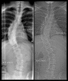
A Comparison of Cobb Angle: Standing Versus Supine Images of Late-Onset Idiopathic Scoliosis
- PMID: 27354881
- PMCID: PMC4912347
- DOI: 10.12659/PJR.895949
A Comparison of Cobb Angle: Standing Versus Supine Images of Late-Onset Idiopathic Scoliosis
Abstract
Background: Scoliosis is traditionally evaluated by measuring the Cobb angle in radiograph images taken while the patient is standing. However, low-dose computed tomography (CT) images, which are taken while the patient is in a supine position, provide new opportunities to evaluate scoliosis. Few studies have investigated how the patient's position, standing or supine, affects measurements. The purpose of this study was to compare the Cobb angle in images from patients while standing versus supine.
Material/methods: A total of 128 consecutive patients (97 females and 21 males; mean age 15.5 [11-26] years) with late-onset scoliosis requiring corrective surgery were enrolled. One observer evaluated the type of curve (Lenke classification) and measured the Cobb angle in whole-spine radiography (standing) and scout images from low-dose CT (supine) were taken on the same day.
Results: For all primary curves, the mean Cobb angle was 59° (SD 12°) while standing and 48° (SD 12°) while in the supine position, with a mean difference of 11° (SD 5°). The correlation between primary standing and supine images had an r value of 0.899 (95% CI 0.860-0.928) and an intra-class correlation coefficient value of 0.969. The correlation between the difference in standing and supine images from primary and secondary curves had an r value of 0.340 (95% CI 0.177-0.484).
Conclusions: We found a strong correlation between the Cobb angle in images obtained while the patient was standing versus supine for primary and secondary curves. This study is only applicable for patients with severe curves requiring surgical treatment. It enables additional studies based on low-dose CT.
Keywords: Scoliosis; Spine; Supine Position.
Figures


Similar articles
-
Supine to standing Cobb angle change in idiopathic scoliosis: the effect of endplate pre-selection.Scoliosis. 2014 Oct 8;9:16. doi: 10.1186/1748-7161-9-16. eCollection 2014. Scoliosis. 2014. PMID: 25342959 Free PMC article.
-
Adult Degenerative Scoliosis: Can Cobb Angle on a Supine Posteroanterior Radiograph Be Used to Predict the Cobb Angle in a Standing Position?Medicine (Baltimore). 2016 Feb;95(6):e2732. doi: 10.1097/MD.0000000000002732. Medicine (Baltimore). 2016. PMID: 26871814 Free PMC article.
-
Supine flexibility predicts curve progression for patients with adolescent idiopathic scoliosis undergoing underarm bracing.Bone Joint J. 2020 Feb;102-B(2):254-260. doi: 10.1302/0301-620X.102B2.BJJ-2019-0916.R1. Bone Joint J. 2020. PMID: 32009436
-
Prediction of Spontaneous Lumbar Curve Correction After Posterior Spinal Fusion for Adolescent Idiopathic Scoliosis Lenke Type 1 Curves.Clin Spine Surg. 2019 Mar;32(2):E112-E116. doi: 10.1097/BSD.0000000000000736. Clin Spine Surg. 2019. PMID: 30379656
-
Comparison of Cobb angles in idiopathic scoliosis on standing radiographs and supine axially loaded MRI.Spine (Phila Pa 1976). 2006 Dec 15;31(26):3039-44. doi: 10.1097/01.brs.0000249513.91050.80. Spine (Phila Pa 1976). 2006. PMID: 17173001
Cited by
-
Comparison of Cobb angle measurements for scoliosis assessment using different imaging modalities: a systematic review.EFORT Open Rev. 2023 Jun 8;8(6):489-498. doi: 10.1530/EOR-23-0032. EFORT Open Rev. 2023. PMID: 37289072 Free PMC article.
-
Impact of customized add-on nighttime bracing in full-time brace treatment of adolescent idiopathic scoliosis.PLoS One. 2023 Jan 26;18(1):e0278421. doi: 10.1371/journal.pone.0278421. eCollection 2023. PLoS One. 2023. PMID: 36701318 Free PMC article.
-
Impact of patient position on coronal Cobb angle measurement in non-ambulatory myelodysplastic patients.Eur J Orthop Surg Traumatol. 2019 Jan;29(1):25-29. doi: 10.1007/s00590-018-2264-1. Epub 2018 Jun 18. Eur J Orthop Surg Traumatol. 2019. PMID: 29915954
References
-
- Cobb JR. Instructional course lectures. The American Academy of Orthopedic Surgeons. 5th ed. Ann Arbor: J. W Edwards; 1948. Outline for the study of scoliosis; pp. 261–75.
-
- Morrissy RT, Goldsmith GS, Hall EC, et al. Measurement of the Cobb angle on radiographs of patients who have scoliosis. Evaluation of intrinsic error. J Bone Joint Surg Am. 1990;72:320–27. - PubMed
-
- Carman DL, Browne RH, Birch JG. Measurement of scoliosis and kyphosis radiographs. Intraobserver and interobserver variation. J Bone Joint Surg Am. 1990;72(3):328–33. - PubMed
-
- Adam CJ, Izatt MT, Harvey JR, et al. Variability in Cobb angle measurements using reformatted computerized tomography scans. Spine. 2005;30(14):1664–69. - PubMed
-
- Perdriolle R, Vidal J. Thoracic idiopathic scoliosis curve evolution and prognosis. Spine. 1985;10(9):785–91. - PubMed
LinkOut - more resources
Full Text Sources
Other Literature Sources
