

Eagle Syndrome Causing Vascular Compression with Cervical Rotation: Case Report
- PMID: 27354882
- PMCID: PMC4912348
- DOI: 10.12659/PJR.896741
Eagle Syndrome Causing Vascular Compression with Cervical Rotation: Case Report
Abstract
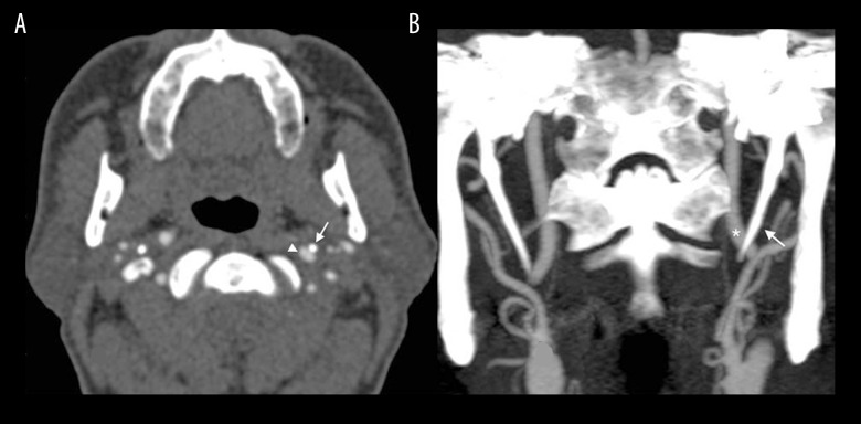
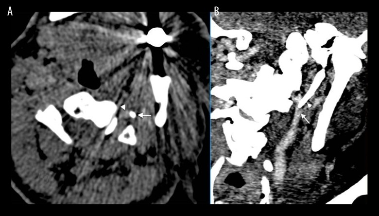
Background: Eagle syndrome is a condition caused by an elongated styloid process. Unilateral face, neck and ear pain, stinging pain, foreign body sensation and dysphagia can be observed with this syndrome. Rarely, the elongated styloid process may cause pain by compressing the cervical segment of the internal carotid and the surrounding sympathetic plexus, and that pain spreading along the artery can cause neurological symptoms such as vertigo and syncope.
Case report: In this case report we presented a very rare eagle syndrome with neurological symptoms that occurred suddenly with cervical rotation. The symptoms disappeared as suddenly as they occurred, with the release of pressure in neutral position. We also discussed CT angiographic findings of this case.
Conclusions: Radiological diagnosis of the Eagle syndrome that is manifested with a wide variety of symptoms and causes diagnostic difficulties when it is not considered in the differential diagnosis is easy in patients with specific findings. CT angiography is a fast and effective examination in terms of showing compression in patients with the Eagle syndrome that is considered to be atypical and causes vascular compression.
Keywords: Angiography; Eagles; Multidetector Computed Tomography; Vertigo.
Figures
References
-
- Gervickas A, Kubilius R, Sabalys G. Clinic, diagnostics and treatment pecularities of Eagle’s syndrome. Stomatologija, Baltic Dental and Maxillofacial Journal. 2004;6:11–13.
-
- Yavuz H, Çakmak Ö, Akkuzu B, Özlüoğlu L. Eagles syndrome: A case report. K.B.B. and BBC Journal. 2002;(10):97–101.
-
- Gök U, Yıldız M. Eagle Sendromu. Fırat Medical Journal. 2004;9(3):79–81.
-
- Hekimoğlu C. Eagle’s Syndrome. Clinical Dentistry and Research. 2005;(3):27–32.
Publication types
LinkOut - more resources
Full Text Sources
Other Literature Sources