. 2016 Mar 21;5(2):e275-80.
doi: 10.1016/j.eats.2016.01.017. eCollection 2016 Apr.

Endoscopy-Assisted Periacetabular Osteotomy

Affiliations

Endoscopy-Assisted Periacetabular Osteotomy

Dean K Matsuda et al. Arthrosc Tech. .

Abstract

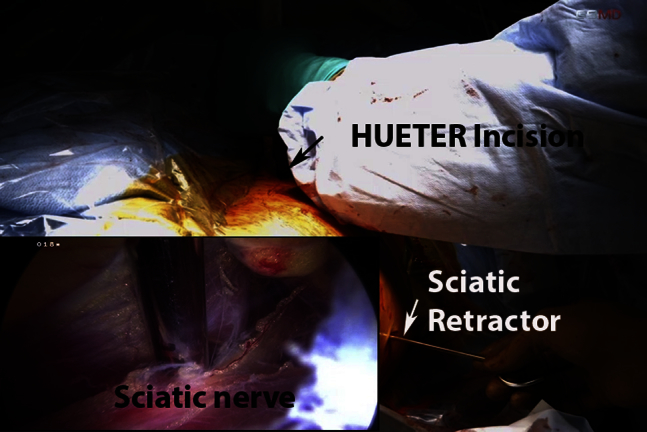
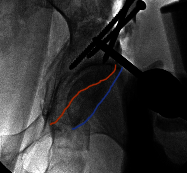
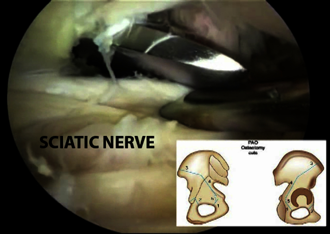
Minimizing soft tissue dissection and improving visualization of vital structures during periacetabular osteotomy (PAO) is of paramount importance to improve patient outcome and minimize complications. The endoscopy-assisted PAO was introduced to accomplish this objective. It involves an initial hip arthroscopy, for treatment of central compartment pathology, followed by a mini-open Bernese periacetabular osteotomy under fluoroscopic and endoscopic guidance, and completed by final dynamic hip arthroscopy to assess acetabular reorientation and fixation and to perform femoroplasty in relation to the new acetabular rim position, if needed. Endoscopy-assisted PAO is used to treat dysplasia or acetabular retroversion in a minimally invasive fashion.


Figures

Fig 1
Fig 1
Representative AP pelvis demonstrating some procedural components of endoscopy-assisted PAO, including intra-articular arthroscopy and extra-articular endoscopy. (AP, anteroposterior; AVN, avascular necrosis; CC dx & rx, central compartment diagnosis and treatment; Femoroplx, femoroplasty; fx, fracture; IA, intra-articular; PAO, periacetabular osteotomy; Post, posterior; THA, total hip arthroplasty.)
Fig 2
Fig 2
Intraoperative photograph of left endoscopy-assisted periacetabular osteotomy in supine position during posterior column osteotomy with osteotome in mini-Hueter incision and endoscopically placed and visualized retractor protecting sciatic nerve (inset) in posterior peritrochanteric space via posterolateral portal as viewed from anterolateral portal with a 70-degree arthroscope.
Fig 3
Fig 3
Intraoperative anteroposterior fluoroscopic image of initial reoriented acetabulum demonstrating large crossover sign above intersection of anterior (red) and posterior (blue) lines. Note K-wires providing transient fixation. Arthroscopy clarified that anterior prominence was caused by anterior inferior iliac spine and not by anterior acetabular rim.
Fig 4
Fig 4
Intraoperative anteroposterior fluoroscopic image of acetabular fragment fixated in a more desirable position with improved anterolateral coverage without pincer or subspine impingement. Note Schanz pin providing acetabular repositioning and fixation with cortical screws. Anterior rim (red line) is medial to the posterior rim (blue line).
Fig 5
Fig 5
Arthroscopic and fluoroscopic (inset) image of left hip from anterolateral portal during femoroplasty after endoscopy-assisted periacetabular osteotomy, enabling optimization of concurrent cam decompression in relation to newly oriented acetabular rim.
Fig 6
Fig 6
Intraoperative dynamic testing confirms eradication of impingement, stability of endoscopy-assisted periacetabular osteotomy fixation, and retention of labral fluid seal (inset as viewed from anterolateral portal) after arthroscopic femoroplasty.
Fig 7
Fig 7
Arthroscopic image of left hip via anterolateral portal showing capsular repair. (C, capsule; FH, femoral head; L, labrum.)
Fig 8
Fig 8
Arthroscopic image of cadaveric left hip demonstrating proximity of osteotome during ischial osteotomy and retraction of adjacent sciatic nerve with blunt obturator. Inset shows the periacetabular osteotomy cuts with ischial osteotomy labeled 1.

References

    1. Ross J.R., Clohisy J.C., Baca G., Sink E., ANCHOR Investigators Patient and disease characteristics associated with hip arthroscopy failure in acetabular dysplasia. J Arthroplasty. 2014;29(9 suppl):160–163. - PubMed
    1. Matsuda D.K., Khatod M. Rapidly progressive osteoarthritis after arthroscopic labral repair in patients with hip dysplasia. Arthroscopy. 2012;28:1738–1743. - PubMed
    1. Fukui K., Briggs K.K., Trindade C.A., Philippon M.J. Outcomes after labral repair in patients with femoroacetabular impingement and borderline dysplasia. Arthroscopy. 2015;31:2371–2379. - PubMed
    1. Domb B.G., Stake C.E., Lindner D., El-Bitar Y., Jackson T.J. Arthroscopic capsular plication and labral preservation in borderline hip dysplasia: Two-year clinical outcomes of a surgical approach to a challenging problem. Am J Sports Med. 2013;41:2591–2598. - PubMed
    1. Ganz R., Klaue K., Vinh T.S., Mast J.W. A new periacetabular osteotomy for the treatment of hip dysplasias. Technique and preliminary results. Clin Orthop Relat Res. 1988;232:26–36. - PubMed

LinkOut - more resources