. 2016 Apr 4;5(2):e337-42.
doi: 10.1016/j.eats.2016.01.005. eCollection 2016 Apr.

Hip Labral Reconstruction: The "Kite Technique" for Improved Efficiency and Graft Control

Affiliations

Hip Labral Reconstruction: The "Kite Technique" for Improved Efficiency and Graft Control

Sanjeev Bhatia et al. Arthrosc Tech. .

Abstract

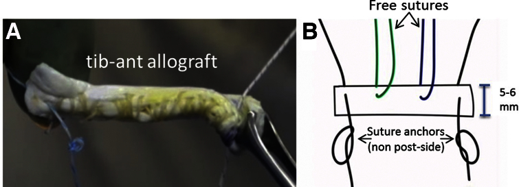
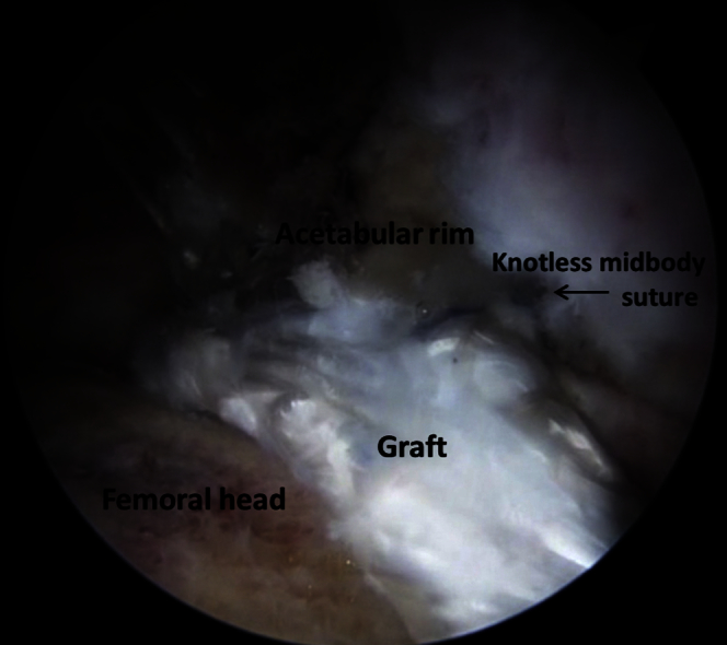
Although the merits of labral reconstruction have been well established, the technical difficulty of presently used reconstruction techniques-particularly with graft passage and fixation-limit its efficacy and potentiates the risk of iatrogenic damage within the hip joint. The unwieldy nature of a floating labral graft anchored on one end may impede accurate fixation of the other end, which is critical for restoration of the fluid hip seal and preservation of graft integrity. In this technique narrative, we present a "kite technique" for introduction, control, and efficient fixation of a labral reconstruction graft. The principles of this method are founded on the belief that a soft-tissue graft in an arthroscopic environment is much easier to guide into position with 2 control sutures using a pulley system similar to flying a kite with 2 fly lines. Although we herein detail the technique as it applies to labral reconstruction in the hip, the concept of the kite technique may also be employed in arthroscopic-assisted soft-tissue reconstructions of other joints.


Figures

Fig 1
Fig 1
Patient positioning. The patient is placed supine and the operative leg (left) is put in traction, adducted, and internally rotated. The perineum and all bony prominences are appropriately padded.
Fig 2
Fig 2
Intraoperative photograph of a left hip showing the location of the portals and their relation to the greater trochanter. (DALA, distal anterolateral accessory portal; MAP, mid-anterior portal; PAL, proximal anterolateral portal.)
Fig 3
Fig 3
A suture anchor is placed at the anterior most aspect of the defect (3 o'clock position is this patient) using the distal anterolateral accessory portal as shown here in an arthroscopic image of a left hip as viewed through the proximal anterolateral portal.
Fig 4
Fig 4
A suture anchor is placed at the posterior most aspect of the defect (11 o'clock position is this patient) using the distal anterolateral accessory portal as shown here in an arthroscopic image of a left hip as viewed through the proximal anterolateral portal.
Fig 5
Fig 5
The prepared graft shown with (A) an intraoperative photograph and (B) an illustrated image showing the post, non-post, and free sutures. The width should be approximately 5-6 mm.
Fig 6
Fig 6
Illustrated image of an alternate option of graft preparation. If added security around the graft is desired for graft passage, the non-post suture can be passed through and around the graft before being tied to itself.
Fig 7
Fig 7
Illustrated diagram of how the graft will be placed to reconstruct the labral defect. The post and non-post sutures are depicted in black and purple, with the square shape indicating the non-post side. In between these sutures are the knotless suture anchors.
Fig 8
Fig 8
Intraoperative image of the graft being inserted through the DALA portal, whereas the arthroscope is in the PAL portal. The graft is inserted into the joint using the “kite technique.” The 2 sutures out of the PAL portal (post sutures) adjacent to the arthroscope are alternatingly tensioned back and forth, similar to fly lines on a kite, and the knots on the ends of the anterior and posterior non-post sutures effectively pull each end of the graft into position along the rim. (DALA, distal anterolateral accessory portal; MAP, mid-anterior portal; PAL, proximal anterolateral portal.)
Fig 9
Fig 9
The anterior most suture is retrieved and tied via the mid-anterior portal.
Fig 10
Fig 10
Knotless mid-body sutures are viewed through the proximal anterolateral portal and retrieved and secured to the acetabular rim through the distal anterolateral accessory portal.
Fig 11
Fig 11
Intraoperative photograph of the finished labral reconstruction viewed through the proximal anterolateral portal.

References

    1. Bedi A., Kelly B.T., Khanduja V. Arthroscopic hip preservation surgery: Current concepts and perspective. Bone Joint J. 2013;95-B:10–19. - PubMed
    1. Philippon M.J., Nepple J.J., Campbell K.J. The hip fluid seal—Part I: The effect of an acetabular labral tear, repair, resection, and reconstruction on hip fluid pressurization. Knee Surg Sports Traumatol Arthrosc. 2014;22:722–729. - PubMed
    1. Nepple J.J., Philippon M.J., Campbell K.J. The hip fluid seal—Part II: The effect of an acetabular labral tear, repair, resection, and reconstruction on hip stability to distraction. Knee Surg Sports Traumatol Arthrosc. 2014;22:730–736. - PubMed
    1. Ferguson S.J., Bryant J.T., Ganz R., Ito K. An in vitro investigation of the acetabular labral seal in hip joint mechanics. J Biomech. 2003;36:171–178. - PubMed
    1. Espinosa N., Rothenfluh D.A., Beck M., Ganz R. Treatment of femoro-acetabular impingement: Preliminary results of labral refixation. J Bone Joint Surg Am. 2006;88:925–935. - PubMed

LinkOut - more resources