Review
. 2016 Sep;89(1065):20160284.
doi: 10.1259/bjr.20160284. Epub 2016 Jun 29.

The MRI features of placental adhesion disorder-a pictorial review

Affiliations
Review

The MRI features of placental adhesion disorder-a pictorial review

Faye Cuthbert et al. Br J Radiol. 2016 Sep.

Abstract

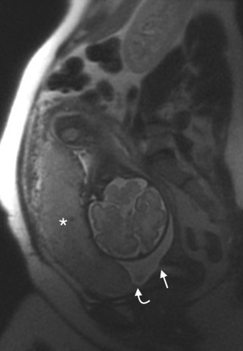
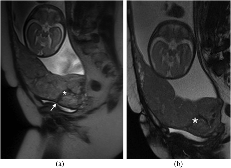
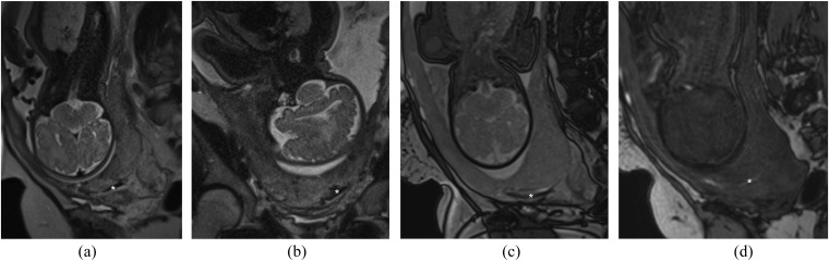
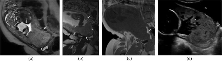
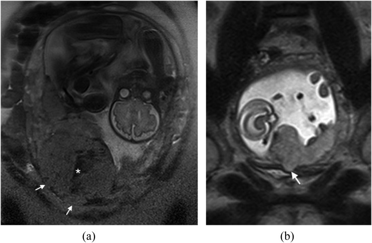
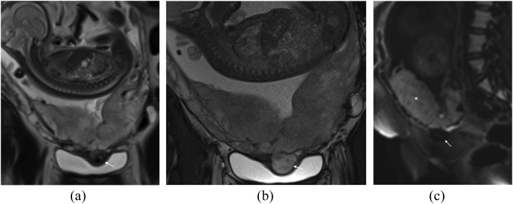
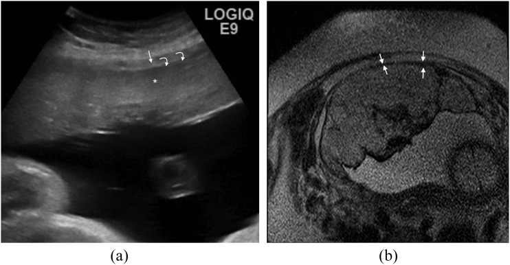
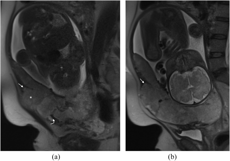
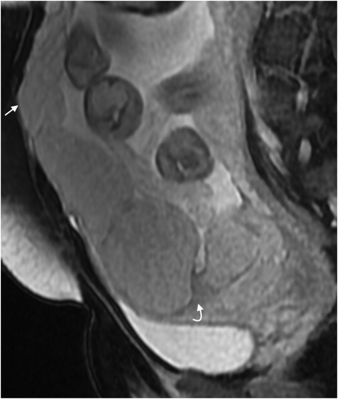
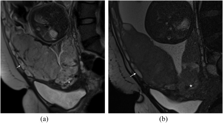
Placental adhesion disorder (PAD) comprises placenta accreta, increta and percreta lesions; these are classified according to the depth of uterine invasion. Although PAD is considered a rare condition, its incidence has increased 10-fold in the last 50 years. Ultrasound is the primary imaging modality for the assessment of the placenta and in the majority of cases, it is sufficient for diagnosis; however, when ultrasound findings are suspicious or inconclusive, MRI is recommended as an adjunct imaging technique. Numerous MRI features of PAD have been described, including dark intraplacental bands, disorganized intraplacental vascularity and abnormal uterine bulging. This pictorial review describes and illustrates these characteristics and discusses their implications in planning delivery. In addition, we present a series of "pitfall" cases to aid the interpreting radiologist and discuss management of PAD. PAD is a clinical and diagnostic challenge that is encountered with increasing frequency, requiring a cohesive multidisciplinary approach to its management.


Figures

Figure 1.
Figure 1.
Normal placenta: sagittal T2 single-shot fast spin-echo image of a normal anterior placenta (asterisk). The leading edge of the placenta (curved arrow) is clearly anterior and superior to the internal cervical os (arrow).
Figure 2.
Figure 2.
Dark intraplacental bands in a placenta percreta: (a) sagittal T2 single-shot fast spin-echo image demonstrates the typical appearance of a dark intraplacental band (asterisk) in a patient with a complete placenta praevia and a prior caesarean section. The caesarian section scar is visible (arrow). (b) Sagittal T2 balanced gradient-echo image demonstrates persistent low T2 signal intensity of the dark intraplacental band (asterisk), this allows differentiation from an abnormal vessel. Histology confirmed a placenta percreta.
Figure 3.
Figure 3.
Pitfall—clot mimicking a dark intraplacental band: (a) sagittal T2 single-shot fast spin-echo (SSFSE) image demonstrates a linear low T2 signal intensity (T2SI) region (asterisk) in the expected region of the previous caesarean section scar. (b) Coronal T2 SSFSE image of the same area, the linear region of low T2SI (asterisk) is again visualized. (c) Sagittal T2 balanced gradient-echo image demonstrates persistent low T2SI (asterisk), suggestive but not typical of a dark intraplacental band. (d) Sagittal T1 weighted image demonstrates this area returns high T1 signal intensity (asterisk) in keeping with haemorrhage. Overall appearances are not in keeping with a dark intraplacental band. At caesarean section, the placenta was removed normally.
Figure 4.
Figure 4.
Disorganized vascularity: (a) sagittal T2 single-shot fast spin-echo demonstrates a complete placenta praevia. The low T2 signal intensity serpiginous vessels along the posterior myometrium are noted (arrow). In addition, the placenta is heterogeneous with a low uterine bulge (curved arrow). (b) Coronal oblique balanced gradient-echo image in the same patient demonstrates extensive disorganized vascularity in the anterior and posterior myometrium (arrows). A uterine bulge (curved arrow) is again noted. (c) Coronal oblique T1 weighted image demonstrates a high T1 signal intensity collection (arrow) in keeping with retroplacental haemorrhage. This case was proven to be a placenta percreta at histology. The appearences of the abnormal vessels should be reported as the information will facilitate surgical planning. (d) Ultrasound image in a different patient demonstrating multiple tortuous hypoechoic structures within the placenta (arrows) in keeping with lacunae. The bladder (B) and the foetus (F) can be noted.
Figure 5.
Figure 5.
Abnormal uterine bulge: (a) coronal T2 single-shot fast spin-echo (SSFSE) image in a patient with placenta percreta. An abnormal uterine bulge can be noted in two areas (arrows). Note also the typical appearance of the dark intraplacental band (asterisk). Placenta percenta was confirmed at histology. (b) Coronal T2 SSFSE image demonstrating an abnormal uterine bulge (arrow) through the previous caesarean section scar in a different patient this scan was acquired at 18/40. The placenta is normal in appearence and this is a case of scar dehiscence not placenta percreta.
Figure 6.
Figure 6.
Pitfall—abnormal uterine bulge: (a) coronal single-shot fast spin-echo image demonstrates a heterogeneous placenta with low T2 signal intensity bulge indenting the bladder (arrow). (b) Coronal T2 balanced gradient-echo image demonstrates the abnormal bulge (arrow) indenting the bladder; it has the same signal intensity as the placenta on these sequences. (c) Sagittal B-800 diffusion-weighted image clearly demonstrates normal restricted diffusion in the placenta (asterisk) but not in the abnormal bulge (arrow). These appearances are consistent with a bladder varix; this proved to be vital information for the obstetrics team to facilitate planning of delivery.
Figure 7.
Figure 7.
Thinning of the myometrium: (a) ultrasound image demonstrating the normal hyperechoic placenta (asterisk) surrounded by the hypoechoic myometrium (arrow). The thin hypoechoic line at the inner aspect of the myometrium (curved arrows) represents the retroplacental clear space. On colour Doppler, a normal organized pattern of subplacental flow that parallels the myometrium is expected. (b) Axial T2 single-shot fast spin-echo image demonstrates thinning of the myometrium (arrows) in a case of placenta percreta. This assessment must be made in three planes and if there is doubt, correlation with ultrasound is often useful.
Figure 8.
Figure 8.
Pitfall—uterine dehiscence: (a) sagittal T2 single-shot fast spin-echo (SSFSE) image demonstrates a dark band in the expected site of the previous caesarean section (curved arrow) and a bulge superiorly (asterisk). This has the appearance of a percreta but at surgery, massive uterine dehiscence was found; the placenta had been “plugging” the breach in the myometrium. Note the myometrium is ending in a “V” shape (arrow), in retrospect in keeping with uterine retraction and dehiscence. (b) Sagittal T2 SSFSE image in the same patient again showing the “V”-shaped uterine retraction (arrow) in keeping with dehiscence.
Figure 9.
Figure 9.
Pitfall—focal bulge in the region of the maternal umbilicus: sagittal T2 balanced gradient-echo image demonstrates a dark band in the low uterine segment at the expected site of the caesarean-section scar (curved arrow); in addition, there is placenta percreta in the region of the cervix. The bulge in the anterior abdominal wall (arrow) could be mistaken for an area of invasion, but it is in fact a common normal finding due to separation of the rectus muscles as pregnancy progresses.
Figure 10.
Figure 10.
Localized area of abnormality: (a) sagittal T2 single-shot fast spin-echo image demonstrates a complete placenta praevia with a solitary low T2 signal intensity linear area in keeping with a dark intraplacental band (asterisk). Note the increased vascularity (arrow) along the anterior myometrium. (b) Sagittal T2 balanced gradient-echo image is confirms the presence of the dark intraplacental band (asterisk) and increased vascularity (arrow). The MRI provided essential pre-delivery information. This placenta came away in a piecemeal fashion with increased blood loss.

Similar articles

Cited by

References

    1. Chantraine F, Blacher S, Berndt S, Palacios-Jaraquemada J, Sarioglu N, Nisolle M, et al. . Abnormal vascular architecture at the placental-maternal interface in placenta increta. Am J Obstet Gynecol 2012; 207: 188.e1–9. doi: 10.1016/j.ajog.2012.06.083 - DOI - PubMed
    1. Eshkoli T, Weintraub AY, Sergienko R, Sheiner E. Placenta accreta: risk factors, perinatal outcomes, and consequences for subsequent births. Am J Obstet Gynecol 2013; 208: 219.e1–7. doi: 10.1016/j.ajog.2012.12.037 - DOI - PubMed
    1. Knight M; UKOSS. Peripartum hysterectomy in the UK: management and outcomes of the associated haemorrhage. BJOG 2007; 114: 1380–7. doi: 10.1111/j.1471-0528.2007.01507.x - DOI - PubMed
    1. Placenta Praevia, Placenta Praevia Accreta and Vasa Praevia: Diagnosis and Management. RCOG (Green-top guideline No. 27). Internet; 2011; 4.6.
    1. NICE, Caesarean Section, in CG132; 2011.