. 2016 Jun 29;11(6):e0158153.
doi: 10.1371/journal.pone.0158153. eCollection 2016.

Regional Volume Decreases in the Brain of Pax6 Heterozygous Mutant Rats: MRI Deformation-Based Morphometry

Affiliations

Regional Volume Decreases in the Brain of Pax6 Heterozygous Mutant Rats: MRI Deformation-Based Morphometry

Kotaro Hiraoka et al. PLoS One. .

Abstract

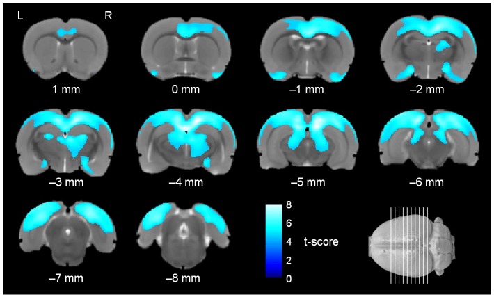
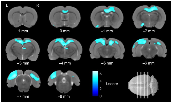
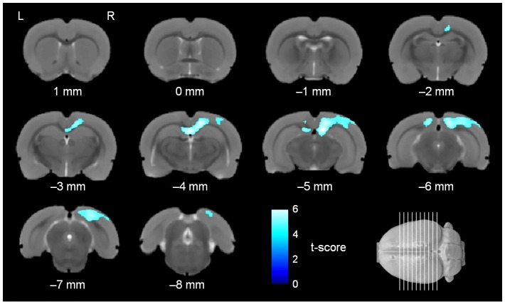
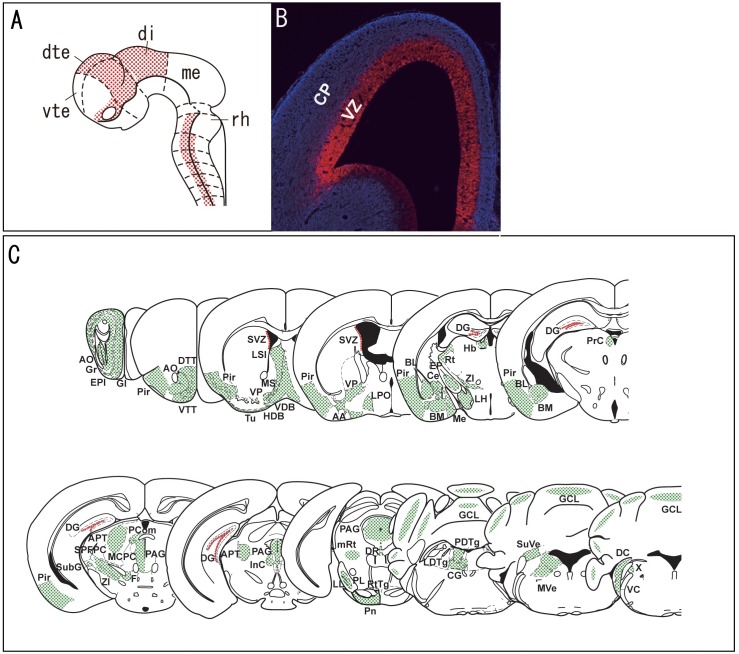
Pax6 is a transcription factor that pleiotropically regulates various developmental processes in the central nervous system. In a previous study, we revealed that Pax6 heterozygous mutant (rSey2/+) adult rats exhibit abnormalities in social interaction. However, the brain malformations underlying the behavioral abnormality are unknown. To elucidate the brain malformations in rSey2/+ rats, we morphometrically analyzed brains of rSey2/+ and wild type rats using small-animal magnetic resonance imaging (MRI). Sixty 10-week-old rats underwent brain MRI (29 rSey2/+ rats and 31 wild type rats). SPM8 software was used for image preprocessing and statistical image analysis. Normalized maps of the Jacobian determinant, a parameter for the expansion and/or contraction of brain regions, were obtained for each rat. rSey2/+ rats showed significant volume decreases in various brain regions including the neocortex, corpus callosum, olfactory structures, hippocampal formation, diencephalon, and midbrain compared to wild type rats. Among brain regions, the anterior commissure showed significant interaction between genotype and sex, indicating the effect of genotype difference on the anterior commissure volume was more robust in females than in males. The rSey2/+ rats exhibited decreased volume in various gray and white matter regions of the brain, which may contribute to manifestation of abnormal social behaviors.


Conflict of interest statement

Competing Interests: The authors have declared that no competing interests exist.

Figures

Fig 1
Fig 1. Cortical regions superimposed on the T2-weighted MRI template.
Insular region is ventral and not shown in the figure.
Fig 2
Fig 2. Regional volume decrease in the brain of rSey2/+ rats compared to WT rats.
Colored voxels represent clusters of significant regional volume decrease in the brain of rSey2/+ rats (n = 29) compared to WT rats (n = 31) in ANOVA (FWE, p < 0.05; threshold of 500 voxels) superimposed on the T2-weighted MRI template. rSey2/+, Pax6 heterozygous mutant; WT, wild-type; ANOVA; analysis of variance; FWE, family-wise error.
Fig 3
Fig 3. Regional volume decrease in the brain of female rSey2/+ rats compared to female WT rats.
Colored voxels represent clusters of significant regional volume decrease in the brain of female rSey2/+ rats (n = 13) compared to female WT rats (n = 15) in the post hoc analysis after ANOVA (FWE, p < 0.05, threshold of 500 voxels), superimposed on the T2-weighted MRI template. rSey2/+, Pax6 heterozygous mutant; WT, wild-type; ANOVA; analysis of variance; FWE, family-wise error.
Fig 4
Fig 4. Regional volume decreases in the brain of male rSey2/+ rats compared to male WT rats.
Colored voxels represent clusters of significant regional volume decreases in the brain of male rSey2/+ rats (n = 16) compared to male WT rats (n = 16) in the post hoc analysis after ANOVA (FWE, p < 0.05, threshold of 500 voxels), superimposed on the T2-weighted MRI template. rSey2/+, Pax6 heterozygous mutant; WT, wild-type; ANOVA; analysis of variance; FWE, family-wise error.
Fig 5
Fig 5
(A) Expression pattern of Pax6 in rat embryo at E10.5. The figure was modified from Kikkawa et al. [50]. dte, dorsal telencephalon; di, diencephalon; me, mesencephalon; rh, rhombencephalon; vte, ventral telencephalon. (B) Pax6 expression in an E14.5 mouse cortex. Pax6 protein (red) is restricted to the ventricular zone (VZ), where neural progenitor cells reside; in contrast, Pax6 is not expressed in the Tuj1-positive (blue) cortical plate (CP). The figure was modified from Osumi and Kikkawa [51]. (C) Expression pattern of Pax6 in the adult mouse based on previously published data (Duan et al., 2013 [41]; Haba et al., 2009 [52]; Kohwi et al., 2005 [53]; Maekawa et al., 2005 [54]). The regions with Pax6 expression in the studies of Duan et al. and Haba et al. are shown in green, and the regions in which Pax6 was expressed in neural stem cells/progenitor cells in the other studies are shown in red. Nomenclature and illustration of various brain regions are based on The Mouse Brain in Stereotaxic Coordinates (2nd Edition) [55]. AA, anterior amygdaloid area; AO, anterior olfactory nucleus; APT, anterior pretectal nucleus; BM/BL, basomedial/basolateral amygdaloid nucleus; Ce, central amygdaloid nucleus; CG, central gray; DG, hippocampal dentate gyrus; DR, dorsal raphe nucleus; EP, entopeduncular nucleus; EPl, external plexiform layer of the olfactory bulb; F, nucleus of the fields of Forel; GCL, granular cell layer of the cerebellum; Gl, glomerular layer of the olfactory bulb; Gr, granule cell layer of the olfactory bulb; Hb, habenular nucleus; I, intercalated nuclei of the amygdala; InC, interstitial nucleus of Cajal; LH, lateral hypothalamic area; LL, nucleus of the lateral lemniscus; LPO, lateral preoptic area; LSI, lateral septal nucleus, intermediate part; MCPC, magnocellular nucleus of the posterior commissure; Me, Medial amygdaloid nucleus; mRt, mesencephalic reticular formation; MS, medial septal nucleus; PAG, periaqueductal gray; PCom, nucleus of the posterior commissure; PDTg/LDTg, posterodorsal/laterodorsal tegmental nucleus; Pir, Piriform cortex; PL, paralemniscal nucleus; Pn, pontine nuclei; PrC, precommissural nucleus; Rt, reticular thalamic nucleus; RtTg, reticulotegmental nucleus of the pons; SubG, subgeniculate nucleus; SuVe/MVe, superior/medial vestibular nucleus; SPFPC, subparafascicular thalamic nucleus, parvicellular part; SVZ, subventricular zone; Tu, olfactory tubercle; VC/DC, ventral/dorsal cochlear nucleus; VDB/HDB, nucleus of the vertical/horizontal limb of the diagonal band; VP, ventral pallidum; VTT/DTT, ventral/dorsal tenia tecta; X, nucleus X; ZI, zona incerta.

References

    1. Osumi N. The role of Pax6 in brain patterning. Tohoku J Exp Med. 2001;193:163–174. - PubMed
    1. Osumi N, Shinohara H, Numayama-Tsuruta K, Maekawa M. Concise review: Pax6 transcription factor contributes to both embryonic and adult neurogenesis as a multifunctional regulator. Stem Cells. 2008;26:1663–1672. 10.1634/stemcells.2007-0884 - DOI - PubMed
    1. Hever AM, Williamson KA, van Heyningen V. Developmental malformations of the eye: the role of PAX6, SOX2 and OTX2. Clin Genet. 2006;69:459–470. - PubMed
    1. Hanson IM. PAX6 and congenital eye malformations. Pediatr Res. 2003;54:791–796. - PubMed
    1. Ton CC, Hirvonen H, Miwa H, Weil MM, Monaghan P, Jordan T, et al. Positional cloning and characterization of a paired box- and homeobox-containing gene from the aniridia region. Cell. 1991;67:1059–1074. - PubMed

Substances