CT Scan of Thirteen Natural Mummies Dating Back to the XVI-XVIII Centuries: An Emerging Tool to Investigate Living Conditions and Diseases in History
- PMID: 27355351
- PMCID: PMC4927149
- DOI: 10.1371/journal.pone.0154349
CT Scan of Thirteen Natural Mummies Dating Back to the XVI-XVIII Centuries: An Emerging Tool to Investigate Living Conditions and Diseases in History
Abstract
Objectives: To correlate the radiologic findings detected with computed tomography scan with anthropological data in 13 naturally mummified bodies discovered during works of recovery of an ancient church in a crypt in Roccapelago, in the Italian Apennines.
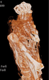
Methods: From a group of about sixty not-intentionally mummified bodies, thirteen were selected to be investigated with volumetric computed tomography (CT). Once CT scan was performed, axial images were processed to gather MPR and Volume Rendering reconstructions. Elaborations of these images provided anthropometric measurements and a non-invasive analysis of the residual anatomical structures. For each body the grade of preservation and the eventual pathological changes were recorded. Furthermore, in order to identify nutritional and occupational markers, radiologic signs of bone tropism and degenerative changes were analysed and graded.
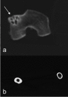
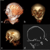
Results: Mummies included seven females and six males, with an estimated age ranging from 20 to 60 years. The first relevant finding identified was a general low grade of preservation, due to the lack of anatomic tissues different from bones, tendons and dehydrated skin. The low grade of preservation was related to the natural process of mummification. Analysing bone degenerative changes on CT scan, the majority of the bodies had significant occupational markers consisting of arthritis in the spine, lower limbs and shoulders even in young age. Few were the pathological findings identified. Among these, the most relevant included a severe bilateral congenital hip dysplasia and a wide osteolytic lesion involving left orbit and petrous bone that was likely the cause of death.
Conclusions: Although the low grade of preservation of these mummies, the multidisciplinary approach of anthropologists and radiologists allowed several important advances in knowledge for the epidemiology of Roccapelago. First of all, a profile of living conditions was delineated. It included occupational and nutritional conditions. Moreover, identification of some causes of death and, most importantly the definition of general living conditions.
Conflict of interest statement
Figures


Similar articles
-
The Sommersdorf mummies-An interdisciplinary investigation on human remains from a 17th-19th century aristocratic crypt in southern Germany.PLoS One. 2017 Aug 31;12(8):e0183588. doi: 10.1371/journal.pone.0183588. eCollection 2017. PLoS One. 2017. PMID: 28859116 Free PMC article.
-
Paleoandrology and prostatic hyperplasia in Italian mummies (XV-XIX century).Med Secoli. 2001;13(2):269-84. Med Secoli. 2001. PMID: 12374108
-
CT checklist and scoring system for the assessment of soft tissue preservation in human mummies: application to catacomb mummies from Palermo, Sicily.Int J Paleopathol. 2018 Mar;20:50-59. doi: 10.1016/j.ijpp.2018.01.003. Epub 2018 Feb 3. Int J Paleopathol. 2018. PMID: 29496216
-
Medical imaging of mummies and bog bodies--a mini-review.Gerontology. 2010;56(5):441-8. doi: 10.1159/000266031. Epub 2009 Dec 11. Gerontology. 2010. PMID: 20016125 Review.
-
What can ancient mummies teach us about atherosclerosis?Trends Cardiovasc Med. 2014 Oct;24(7):279-84. doi: 10.1016/j.tcm.2014.06.005. Epub 2014 Jul 3. Trends Cardiovasc Med. 2014. PMID: 25106086 Review.
Cited by
-
Giant cell tumor of bone in an eighteenth-century Italian mummy.Virchows Arch. 2021 Dec;479(6):1255-1261. doi: 10.1007/s00428-021-03192-5. Epub 2021 Aug 30. Virchows Arch. 2021. PMID: 34462806 Free PMC article.
-
The Sommersdorf mummies-An interdisciplinary investigation on human remains from a 17th-19th century aristocratic crypt in southern Germany.PLoS One. 2017 Aug 31;12(8):e0183588. doi: 10.1371/journal.pone.0183588. eCollection 2017. PLoS One. 2017. PMID: 28859116 Free PMC article.
-
Evidence of neurofibromatosis type 1 in a multi-morbid Inca child mummy: A paleoradiological investigation using computed tomography.PLoS One. 2017 Apr 12;12(4):e0175000. doi: 10.1371/journal.pone.0175000. eCollection 2017. PLoS One. 2017. PMID: 28403237 Free PMC article.
-
Study of a seventeenth-century French artificial mummy: autopsical, native, and contrast-injected CT investigations.Int J Legal Med. 2018 Sep;132(5):1405-1413. doi: 10.1007/s00414-018-1830-8. Epub 2018 Mar 28. Int J Legal Med. 2018. PMID: 29594348
-
The Role of 3D Virtual Anatomy and Scanning Environmental Electron Microscopy in Understanding Morphology and Pathology of Ancient Bodies.Tomography. 2025 Jan 3;11(1):5. doi: 10.3390/tomography11010005. Tomography. 2025. PMID: 39852685 Free PMC article.
References
-
- Cesarani F, Martina MC, Ferraris A, Grilletto R, Boano R, Marochetti EF et al. Whole-Body Three-Dimensional Multidetector CT of 13 Egyptian Human Mummies. Am J Roentgenol 2003; 180: 597–606. - PubMed
-
- Chhem RK, Brothwell DR. Paleoradiology Imaging mummies and fossils. Berlin: Springer; 2008.
-
- Chhem R, Ruhli FJ. Paleoradiology: current status and future challenges. Can Assoc Radiol J 2004; 55: 198–99. - PubMed
-
- Ruhli FJ, Chhem R, Boni T. Diagnostic paleoradiology of mummified tissue: interpretation and pitfalls. Can Assoc Radiol J. 2004; 55: 218–227. - PubMed
Publication types
MeSH terms
LinkOut - more resources
Full Text Sources
Other Literature Sources
Medical

