. 2016 Jun 29;11(6):e0158417.
doi: 10.1371/journal.pone.0158417. eCollection 2016.

Generation of Anti-Boa Immunoglobulin Antibodies for Serodiagnostic Applications, and Their Use to Detect Anti-Reptarenavirus Antibodies in Boa Constrictor

Affiliations

Generation of Anti-Boa Immunoglobulin Antibodies for Serodiagnostic Applications, and Their Use to Detect Anti-Reptarenavirus Antibodies in Boa Constrictor

Yegor Korzyukov et al. PLoS One. .

Abstract

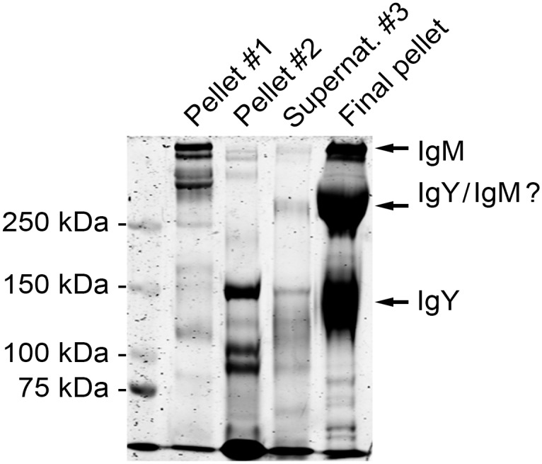
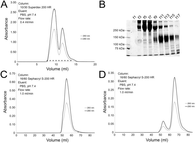
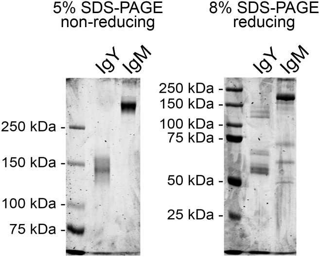
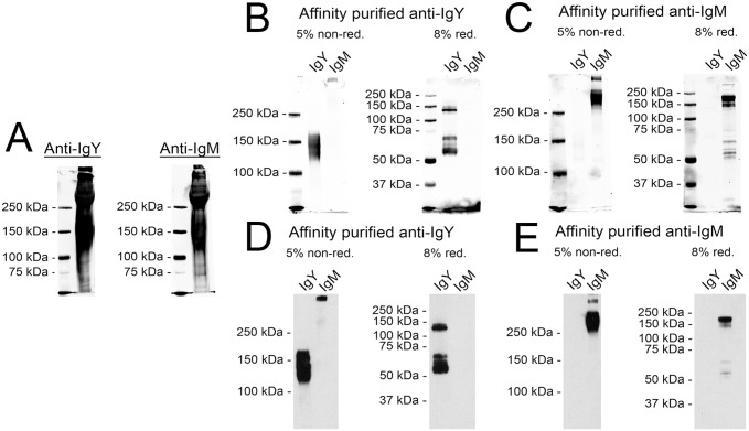
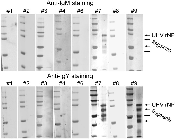
Immunoglobulins (Igs), the key effectors of the adaptive immune system, mediate the specific recognition of foreign structures, i.e. antigens. In mammals, IgM production commonly precedes the production of IgG in the response to an infection. The reptilian counterpart of IgG is IgY, but the exact kinetics of the reptilian immune response are less well known. Boid inclusion body disease (BIBD), an often fatal disease of captive boas and pythons has been linked to reptarenavirus infection, and BIBD is believed to be immunosuppressive. However, so far, the study of the serological response towards reptarenaviruses in BIBD has been hampered by the lack of reagents. Thus we set up a purification protocol for boa constrictor IgY and IgM, which should also be applicable for other snake species. We used centrifugal filter units, poly ethylene glycol precipitation and gel permeation chromatography to purify and separate the IgM and IgY fractions from boa constrictor serum, which we further used to immunise rabbits. We affinity purified IgM and IgY specific reagents from the produced antiserum, and labelled the reagents with horseradish peroxidase. Finally, using the sera of snakes with known exposure to reptarenaviruses we demonstrated that the newly generated reagents can be utilised for serodiagnostic purposes, such as immunoblotting and immunofluorescent staining. To our knowledge, this is the first report to show reptarenavirus-specific antibodies in boa constrictors.


Conflict of interest statement

Competing Interests: The authors have declared that no competing interests exist.

Figures

Fig 1
Fig 1. Polyethylene glycol (PEG) precipitation of proteins from B. constrictor serum.
The serum proteins were initially transferred into PBS using a 100 kDa cut off centrifugal filter device, and precipitation was achieved by step-wise addition of PEG followed by centrifugation. The pelleted proteins were analysed by SDS-PAGE under non-reducing conditions. The left lane shows the molecular weight marker (Precision Plus Protein Dual Color, Bio-Rad), the second lane shows protein precipitated at ~3.5% PEG (pellet #1), the third lane shows protein precipitated at ~12% PEG (supernatant #2), the fourth lane shows protein in supernatant after the second pelleting (supernatant #3), and the fifth lane shows proteins precipitated at ~15% PEG (final pellet).
Fig 2
Fig 2. Purification of IgM and IgY using gel permeation chromatography.
A) The IgM and IgY containing pellet from the PEG precipitation was loaded onto a 10/30 Superdex 200HR column (GE Healthcare) and the proteins were eluted with PBS at a flow rate of 0.4 ml/min. Dual wave length absorbance monitoring, A260nm and A280nm, was used for protein detection. B) Fractions collected during the gel permeation chromatography were analysed by SDS-PAGE under non-reducing conditions, the left lane shows molecular weight marker (Precision Plus Protein Dual Color, Bio-Rad) and the subsequent lanes labelled f1-f17 represent the fractions (marked with * in the chromatogram). C) The fractions (the left “peak” in A, corresponding to f1-f9 in B) containing IgM were pooled, concentrated using a 100 kDa centrifugal filter (Millipore), and loaded onto a 16/60 Sephacryl S-200HR column (GE Healthcare). The proteins were eluted with PBS at flow rate of 1.0 ml/min, and dual wave length absorbance monitoring, A260nm and A280nm, was used for protein detection. D) The fractions (the second “peak” in A, corresponding to f10-f17 in B) containing IgY were pooled, concentrated using a 100 kDa centrifugal filter (Millipore), and loaded onto a 16/60 Sephacryl S-200HR column (GE Healthcare). The proteins were eluted with PBS at a flow rate of 1.0 ml/min, and dual wave length absorbance monitoring, A260nm and A280nm, was used for protein detection.
Fig 3
Fig 3. SDS-PAGE analysis of the purified IgM and IgY.
The IgM and IgY containing fractions were concentrated using a 100 kDa cut off centrifugal filter (Millipore), and the protein composition determined by SDS-PAGE. The left panel shows proteins separated and visualised using Coomassie staining on a 5% SDS-PAGE under non-reducing conditions, the right panel shows proteins separated on a 8% SDS-PAGE under reducing conditions. The left lane in both gels shows the molecular weight marker (Precision Plus Protein Dual Color, Bio-Rad).
Fig 4
Fig 4. Characterisation and purification of the antisera against IgM and IgY.
A) The antisera produced by immunizing rabbits with either purified IgY (Anti-IgY) or IgM (Anti-IgM) were used for immunoblotting the proteins present in the final pellet of the PEG precipitation (see lane 5 in Fig 1 for reference). The left lane in both gels shows the molecular weight marker (Precision Plus Protein Dual Color, Bio-Rad). The results were recorded using Odyssey infrared imaging system (LI-COR Biosciences). B) Immunoblots with affinity purified anti-IgY. The left lanes show the molecular weight marker (Precision Plus Protein Dual Color, Bio-Rad), the middle lanes represent the purified IgY fraction, and the right lanes the purified IgM fraction (see Fig 3 for IgY and IgM reference). The proteins were separated on both 5% SDS-PAGE under non-reducing and 8% SDS-PAGE under reducing conditions. The results were recorded using Odyssey infrared imaging system (LI-COR Biosciences). C) Immunoblots with affinity purified anti-IgM. The lanes and the detection are as in B. D) Immunoblots with affinity purified and HRP labelled anti-IgY. The left lanes represent the purified IgY fraction, and the right lanes the purified IgM fraction. The proteins were separated on both 5% SDS-PAGE under non-reducing and 8% SDS-PAGE under reducing conditions. The marker is not visible, since the results were recorded on X-ray film using enhanced chemiluminescence. E) Immunoblots with affinity purified and HRP labelled anti-IgM. The lanes and the detection are as in D.
Fig 5
Fig 5. Screening of sera from Boa constrictors with BIBD for antibodies against reptarenaviruses by immunoblotting.
Protein pools of Ni-NTA affinity purified recombinant UHV NP (the full-length protein, and N- and C-terminal fragments) separated by SDS-PAGE under reducing conditions were used as the antigens. The membranes were probed with snake serum, and detection of binding was done using either anti-IgY or anti-IgM in combination with an IRDye800 labelled secondary antibody. The immunoblot signals were recorded with Odyssey infrared imaging system (LI-COR Biosciences). The snake serum (or blood) samples #1–4 to #6–9 correspond to the animal numbers of our earlier study (see Table 1, and [20]). The left lanes show the molecular weight marker (Precision Plus Protein Dual Color, Bio-Rad). The arrows indicate the size of the recombinant proteins.
Fig 6
Fig 6. Immunofluorescence staining of reptarenavirus infected cells using protein specific antibodies and snake sera.
A) Vero E6 cells infected with UHV adapted to grow in these cells were fixed at 2 dpi and stained with antibodies against NP (anti-NP-N and anti-NP-C), Z protein (anti-ZP), UHV (anti-UHV). B) The left panels: mock infected Vero E6 cells stained with snake serum #7, which was found positive for both IgM and IgY in immunoblot (see Fig 5), the panels on right: Vero E6 cells infected with UHV (as in A) and stained with snake serum #6, which was found negative for both IgM and IgY in immunoblot (see Fig 5). C) Vero E6 cells infected with UHV (as in A) stained with snake sera #7 (both IgM and IgY positive in immunoblot, Fig 5) and #9 (IgM negative and IgY positive in immunoblot, Fig 5). The antibody binding was detected using both anti-IgY and anti-IgM antibodies and AlexaFluor488 conjugated donkey anti-rabbit immunoglobulins (Molecular Probes). Nuclei were visualized using Hoechst 33342.

References

    1. Schroeder HW Jr, Cavacini L. Structure and function of immunoglobulins. J Allergy Clin Immunol. 2010;125:S41–52. 10.1016/j.jaci.2009.09.046 - DOI - PMC - PubMed
    1. Litman GW, Anderson MK, Rast JP. Evolution of antigen binding receptors. Annu Rev Immunol. 1999. 17:109–147. - PubMed
    1. Zimmerman LM, Vogel LA, Bowden RM. Understanding the vertebrate immune system: Insights from the reptilian perspective. J Exp Biol. 2010. 213(5):661–671. 10.1242/jeb.038315 - DOI - PubMed
    1. Kluger MJ, Ringler DH, Anver MR. Fever and survival. Science. 1975. 188(4184):166–168. - PubMed
    1. Jaffredo T, Fellah JS, and Dunon D. Immunology of Birds and Reptiles. eLS. 2006. 10.1038/npg.els.0000521 - DOI

LinkOut - more resources