. 2016 Jun 29;11(6):e0158358.
doi: 10.1371/journal.pone.0158358. eCollection 2016.

HIF-1alpha Deficiency Attenuates the Cardiomyogenesis of Mouse Embryonic Stem Cells

Affiliations

HIF-1alpha Deficiency Attenuates the Cardiomyogenesis of Mouse Embryonic Stem Cells

Jana Kudová et al. PLoS One. .

Abstract

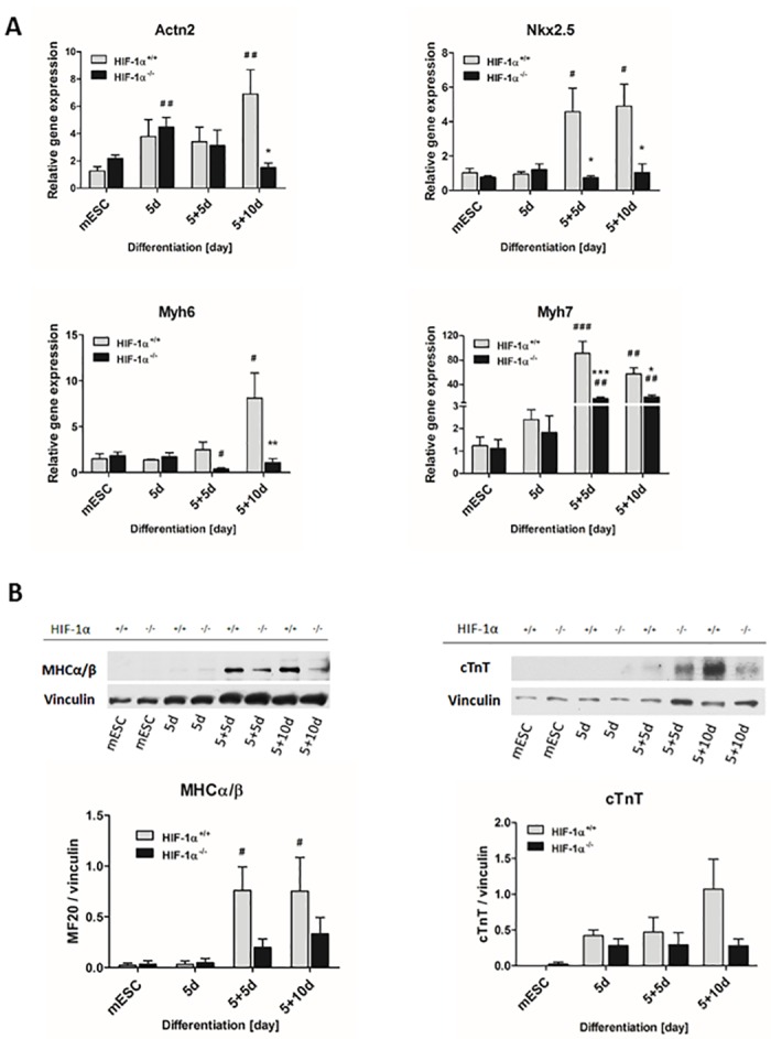
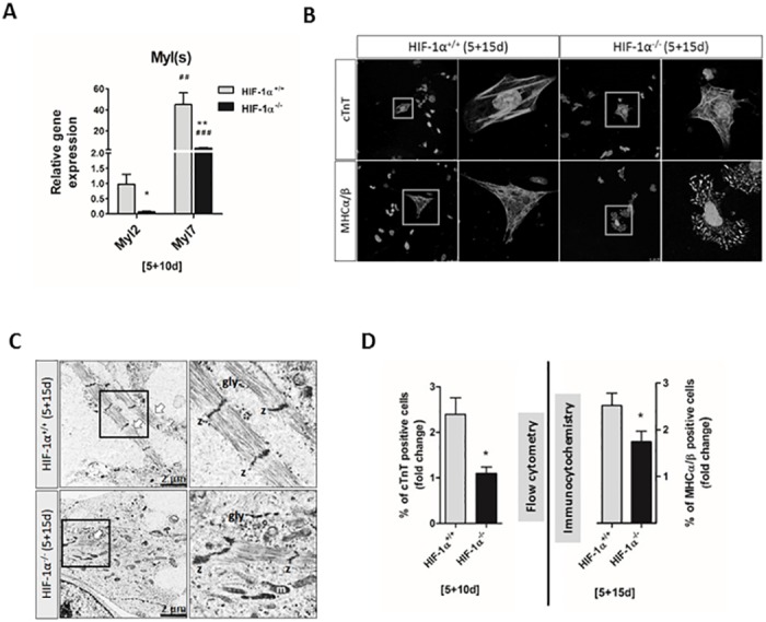
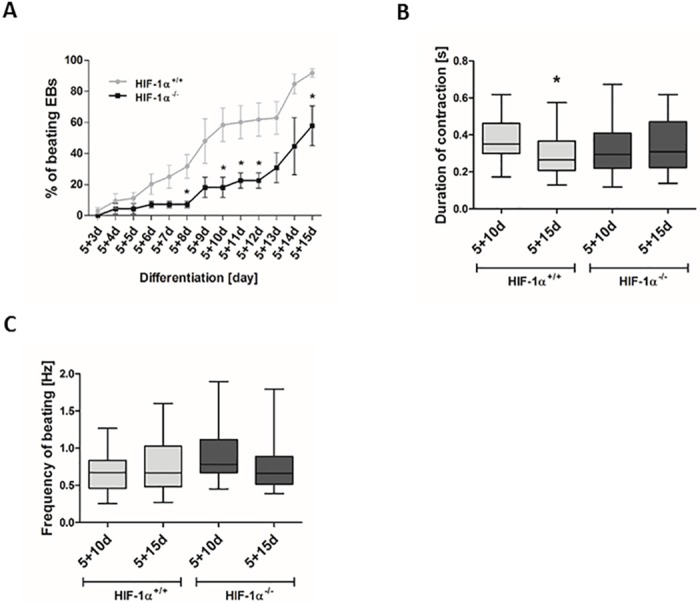
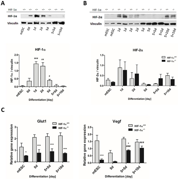
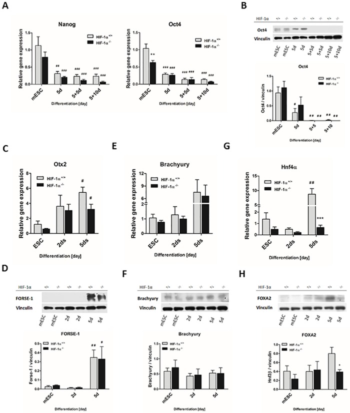
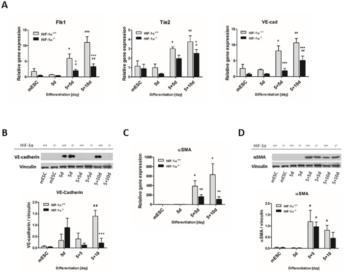
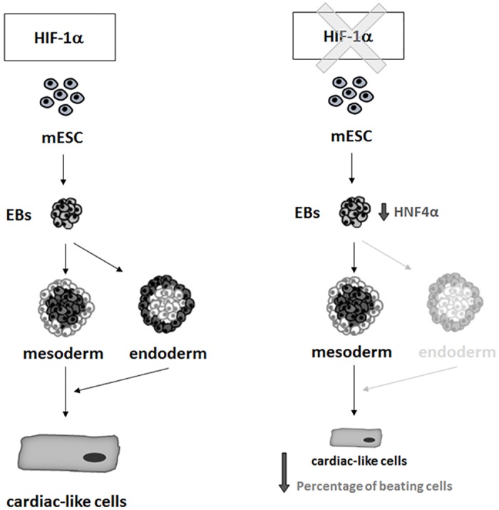
Cardiac cell formation, cardiomyogenesis, is critically dependent on oxygen availability. It is known that hypoxia, a reduced oxygen level, modulates the in vitro differentiation of pluripotent cells into cardiomyocytes via hypoxia inducible factor-1alpha (HIF-1α)-dependent mechanisms. However, the direct impact of HIF-1α deficiency on the formation and maturation of cardiac-like cells derived from mouse embryonic stem cells (mESC) in vitro remains to be elucidated. In the present study, we demonstrated that HIF-1α deficiency significantly altered the quality and quantity of mESC-derived cardiomyocytes. It was accompanied with lower mRNA and protein levels of cardiac cell specific markers (myosin heavy chains 6 and 7) and with a decreasing percentage of myosin heavy chain α and β, and cardiac troponin T-positive cells. As to structural aspects of the differentiated cardiomyocytes, the localization of contractile proteins (cardiac troponin T, myosin heavy chain α and β) and the organization of myofibrils were also different. Simultaneously, HIF-1α deficiency was associated with a lower percentage of beating embryoid bodies. Interestingly, an observed alteration in the in vitro differentiation scheme of HIF-1α deficient cells was accompanied with significantly lower expression of the endodermal marker (hepatic nuclear factor 4 alpha). These findings thus suggest that HIF-1α deficiency attenuates spontaneous cardiomyogenesis through the negative regulation of endoderm development in mESC differentiating in vitro.


Conflict of interest statement

Competing Interests: The authors have declared that no competing interests exist.

Figures

Fig 1
Fig 1. Schematic illustration of the protocol used for the in vitro differentiation of mESC mESC- mouse embryonic stem cells; DMEM- Dulbecco’s modified Eagle’s medium; LIF- leukemia inhibitory factor; FBS- fetal bovine serum; ITS- insulin-transferrin-selenium.
Fig 2
Fig 2. Effect of HIF-1α deficiency on cardiomyogenesis in vitro.
(A) Relative gene expression of selected cardiac markers detected at mRNA level during cardiomyogenesis of wild type and HIF-1α-deficient mESC as revealed by real-time quantitative PCR analysis. The mRNA levels of α-actinin (Actn2), NK2 transcription factor related locus 5 (Nkx2.5), myosin heavy chain 6 (Myh6), and myosin heavy chain 7 (Myh7) were analyzed in undifferentiated ESC (mESC) and in 5-day-old EBs (5d) further differentiated for 5 or 10 days (5+5d, 5+10d). Data are presented as means ± SEM from at least 5 independent experiments; * p < 0.05, ** p < 0.01, *** p < 0.001 (HIF-1α+/+ vs. HIF-1α-/- cells); # p < 0.05, ## p < 0.01, ### p < 0.001 (compared to parental mESC). (B) Protein levels of myosin heavy chains α and β (MHCα/β detected using anti-MF20 antibody) and cardiac troponin T were detected by western blot in the cells derived from wild type and HIF-1α deficient mESC and normalized to the vinculin signal. The densitometric analysis was performed on the basic of 4 independent experiments and data are presented as means ± SEM, # p < 0.05 (compared to parental mESC).
Fig 3
Fig 3. Characterization of wild type and HIF-1α deficient mESC-CMs.
(A) The mRNA levels of myosin light chain 2 and 7 (Myl2, Myl7) genes as revealed by real-time quantitative PCR analysis. Data are presented as means ± SEM from at least 4 independent experiments; * p < 0.05, ** p < 0.01 (HIF-1α+/+ vs. HIF-1α-/- cells); ## p < 0.01, ### p < 0.001 (compared to parental mESC). (B) The intracellular localization of cardiac troponin T (cTnT) and myosin heavy chain α and β (MHCα/β-using anti-MF20 antibody) proteins was specified by confocal microscopy in wild type and HIF-1α deficient differentiated cells (5+15d) processed from at least 3 independent experiments. (C) Transmission electron microscopy of wild type and HIF-1α deficient mESC-CMs at 5+15 day. Data are presented as means ± SEM from 4 independent experiments. The arrows indicate the presence of myofibrils, z Z-disk, gly glycogen and m mitochondria. (D) Analysis of the percentage of cardiac troponin T- positive cells (5+10d) performed by flow cytometry, and of myosin heavy chain α and β- (MHCα/β-using anti-MF20 antibody) positive cells (5+15d) performed by immunocytochemistry. Data are presented as means ± SEM of HIF-1α+/+ cell fold change from at least 3 independent experiments; * p < 0.05 (HIF-1α+/+ vs. HIF-1α-/- cells).
Fig 4
Fig 4. Effect of HIF-1α deficiency on the generation of beating embryonic bodies and cardiac cells.
(A) Beating areas within EBs were counted from day 8 of differentiation (5+3d), (B) the duration of contraction and (C) frequency of beating was analyzed in both 5+10d, 5+15d cells and determined using CBAnalyser. Data are presented as means ± SEM from 3 independent experiments.
Fig 5
Fig 5. Stabilization and transcriptional activity of HIFs during in vitro differentiation.
The protein levels of (A) HIF-1α and (B) HIF-2α were evaluated in wild type and HIF-1α deficient cells by western blot and normalized to the vinculin signal. Data are presented as means ± SEM. The densitometric analysis is representative of 3 independent experiments; # p < 0.05, ## p < 0.01, ### p < 0.001 (compared to parental mESC). (C) The mRNA levels of HIF-1α target genes glucose transporter 1 (Glut1) and vascular endothelial growth factor (Vegf) were measured by real-time quantitative PCR analysis. Data are presented as mean ± SEM from at least 4 independent experiments; * p < 0.05, *** p < 0.001 (HIF-1α+/+ vs. HIF-1α-/- cells); ### p < 0.001 (compared to parental mESC).
Fig 6
Fig 6. Effect of HIF-1α deficiency on lineage specification of spontaneously differentiating mESC.
The mRNA levels of (A) embryonic stem cell markers Nanog and octamer-binding transcription factor 4 (Oct4); (C) ectoderm marker orthodenticle homolog 2 (Otx2); (E) mesoderm marker Brachyury; and (G) endoderm marker hepatic nuclear factor 4 α (HNF4α) were evaluated in wild type and HIF-1α deficient cells during the early stage of differentiation (mESC, 2d, 5d) using real-time quantitative PCR analysis. Data are presented as means ± SEM from at least 3 independent experiments; *** p < 0.001 (HIF-1α+/+ vs. HIF-1α-/- cells); # p < 0.05, ## p < 0.01 (compared to parental mESC). The protein levels of (B) Oct4, (D) FORSE-1, (F) Brachyury and (H) Forkhead Box A2 (FOXA2) were determined by western blot analysis in the cells derived from wild type and HIF-1α deficient mESC and normalized to the vinculin signal. The densitometric analysis was performed on the basic of 3 independent experiments and data are presented as means ± SEM, * p < 0.05 (HIF-1α+/+ vs. HIF-1α-/- cells); # p < 0.05, ## p < 0.01 (compared to parental mESC).
Fig 7
Fig 7. Effect of HIF-1α deficiency on cellular fate of spontaneously differentiating mESC.
The gene expression profiles of (A) endothelial differentiation markers and (C) smooth muscle marker were analyzed during the differentiation process of wild type and HIF-1α deficient cells. The mRNA levels of (A) TEK tyrosine kinase (Tie2), VE-cadherin (VE-cad) and Fetal Liver Kinase 1 (Flk1) and (C) alpha smooth muscle actin (αSMA) were measured by real-time quantitative PCR analysis. Data are presented as means ± SEM from at least 5 independent experiments; * p < 0.05, ** p < 0.01, *** p < 0.001 (HIF-1α+/+ vs. HIF-1α-/- cells); # p < 0.05, ## p < 0.01, ### p < 0.001 (compared to parental mESC). The protein levels of (B) VE-cadherin and (D) αSMA were determined by western blot analysis in the cells derived from wild type and HIF-1α deficient mESC and normalized to the vinculin signal. The densitometric analysis was performed on the basic of 3 independent experiments and data are presented as means ± SEM, *** p < 0.001 (HIF-1α+/+ vs. HIF-1α-/- cells); # p < 0.05, ## p < 0.01 (compared to parental mESC).
Fig 8
Fig 8. Scheme indicating the possible role of HIF-1α in the regulation of cardiomyogenesis in vitro.
For more details see discussion.

Similar articles

Cited by

References

    1. Braunwald E, Bristow MR. Congestive heart failure: fifty years of progress. Circulation. 2000;102(20 Suppl 4):IV14–23. Epub 2000/11/18. . - PubMed
    1. Krishnan J, Ahuja P, Bodenmann S, Knapik D, Perriard E, Krek W, et al. Essential role of developmentally activated hypoxia-inducible factor 1alpha for cardiac morphogenesis and function. Circ Res. 2008;103(10):1139–46. Epub 2008/10/14. 10.1161/01.RES.0000338613.89841.c1 . - DOI - PubMed
    1. Medley TL, Furtado M, Lam NT, Idrizi R, Williams D, Verma PJ, et al. Effect of oxygen on cardiac differentiation in mouse iPS cells: role of hypoxia inducible factor-1 and Wnt/beta-catenin signaling. PLoS One. 2013;8(11):e80280 Epub 2013/11/23. 10.1371/journal.pone.0080280 - DOI - PMC - PubMed
    1. Majmundar AJ, Wong WJ, Simon MC. Hypoxia-inducible factors and the response to hypoxic stress. Mol Cell. 2010;40(2):294–309. Epub 2010/10/23. 10.1016/j.molcel.2010.09.022 - DOI - PMC - PubMed
    1. Hu CJ, Wang LY, Chodosh LA, Keith B, Simon MC. Differential roles of hypoxia-inducible factor 1alpha (HIF-1alpha) and HIF-2alpha in hypoxic gene regulation. Mol Cell Biol. 2003;23(24):9361–74. Epub 2003/12/04. - PMC - PubMed

MeSH terms