

Generation and Feasibility Assessment of a New Vehicle for Cell-Based Therapy for Treating Corneal Endothelial Dysfunction
- PMID: 27355373
- PMCID: PMC4927169
- DOI: 10.1371/journal.pone.0158427
Generation and Feasibility Assessment of a New Vehicle for Cell-Based Therapy for Treating Corneal Endothelial Dysfunction
Abstract
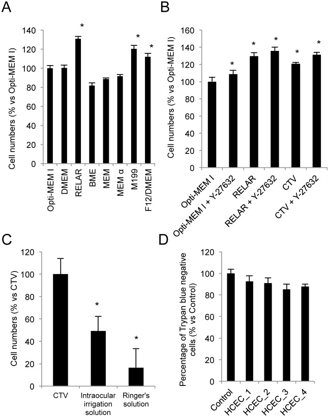
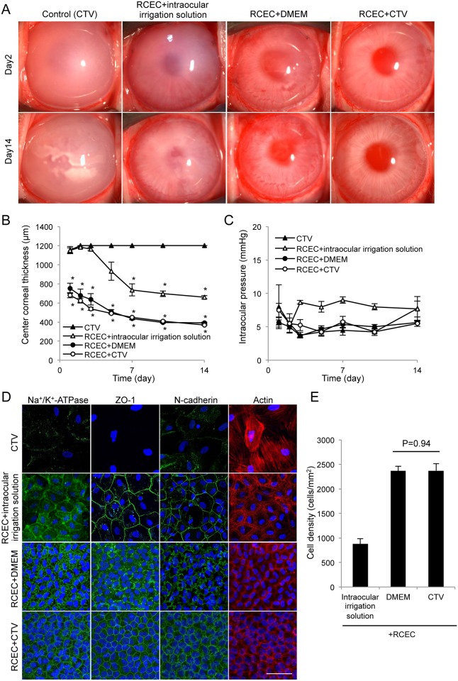
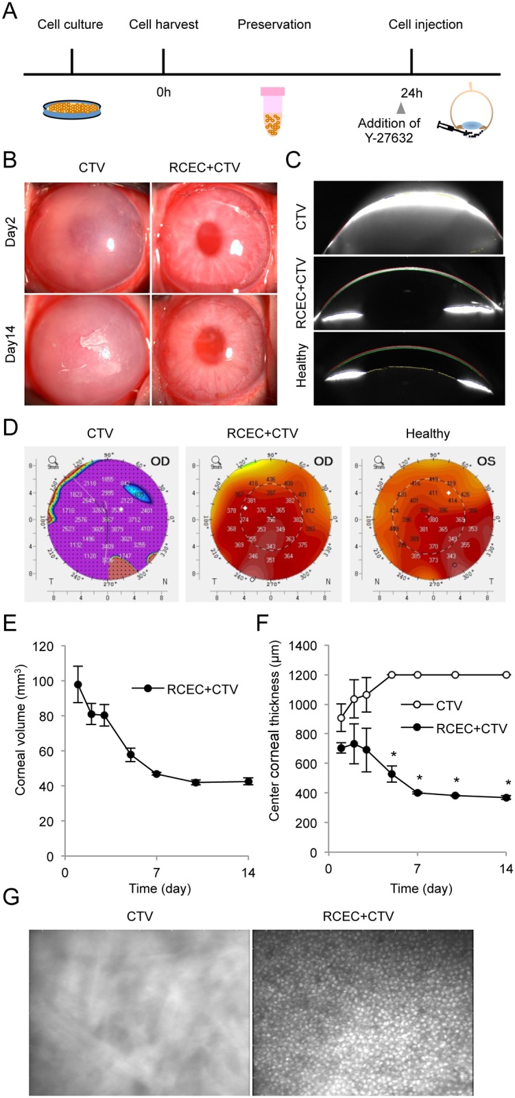
The corneal endothelium maintains corneal transparency by its pump and barrier functions; consequently, its decompensation due to any pathological reason causes severe vision loss due to corneal haziness. Corneal transplantation is the only therapeutic choice for treating corneal endothelial dysfunction, but associated problems, such as a shortages of donor corneas, the difficulty of the surgical procedure, and graft failure, still need to be resolved. Regenerative medicine is attractive to researchers as a means of providing innovative therapies for corneal endothelial dysfunction, as it now does for other diseases. We previously demonstrated the successful regeneration of corneal endothelium in animal models by injecting cultured corneal endothelial cells (CECs) in combination with a Rho kinase (ROCK) inhibitor. The purpose of the present study was to optimize the vehicle for clinical use in cell-based therapy. Our screening of cell culture media revealed that RELAR medium promoted CEC adhesion. We then modified RELAR medium by removing hormones, growth factors, and potentially toxic materials to generate a cell therapy vehicle (CTV) composed of amino acid, salts, glucose, and vitamins. Injection of CECs in CTV enabled efficient engraftment and regeneration of the corneal endothelium in the rabbit corneal endothelial dysfunction model, with restoration of a transparent cornea. The CECs retained >85% viability after a 24 hour preservation as a cell suspension in CTV at 4°C and maintained their potency to regenerate the corneal endothelium in vivo. The vehicle developed here is clinically applicable for cell-based therapy aimed at treating the corneal endothelium. Our strategy involves the generation of vehicle from a culture medium appropriate for a given cell type by removing materials that are not favorable for clinical use.
Conflict of interest statement
Figures

Similar articles
-
ROCK inhibitor converts corneal endothelial cells into a phenotype capable of regenerating in vivo endothelial tissue.Am J Pathol. 2012 Jul;181(1):268-77. doi: 10.1016/j.ajpath.2012.03.033. Epub 2012 Jun 14. Am J Pathol. 2012. PMID: 22704232
-
The Role of Rho Kinase Inhibitors in Corneal Endothelial Dysfunction.Curr Pharm Des. 2017;23(4):660-666. doi: 10.2174/1381612822666161205110027. Curr Pharm Des. 2017. PMID: 27917718 Review.
-
The new therapeutic concept of using a rho kinase inhibitor for the treatment of corneal endothelial dysfunction.Cornea. 2011 Oct;30 Suppl 1:S54-9. doi: 10.1097/ICO.0b013e3182281ee1. Cornea. 2011. PMID: 21912232
-
Rho kinase inhibitor enables cell-based therapy for corneal endothelial dysfunction.Sci Rep. 2016 May 18;6:26113. doi: 10.1038/srep26113. Sci Rep. 2016. PMID: 27189516 Free PMC article.
-
Cell-based approach for treatment of corneal endothelial dysfunction.Cornea. 2014 Nov;33 Suppl 11:S37-41. doi: 10.1097/ICO.0000000000000229. Cornea. 2014. PMID: 25188790 Review.
Cited by
-
Impact of the clinical use of ROCK inhibitor on the pathogenesis and treatment of glaucoma.Jpn J Ophthalmol. 2018 Mar;62(2):109-126. doi: 10.1007/s10384-018-0566-9. Epub 2018 Feb 14. Jpn J Ophthalmol. 2018. PMID: 29445943 Review.
-
Ex vivo cultivated retinal pigment epithelial cell transplantation for the treatment of rabbit corneal endothelial dysfunction.Eye Vis (Lond). 2023 Aug 2;10(1):34. doi: 10.1186/s40662-023-00351-4. Eye Vis (Lond). 2023. PMID: 37528478 Free PMC article.
-
A Framework for Human Corneal Endothelial Cell Culture and Preliminary Wound Model Experiments with a New Cell Tracking Approach.Int J Mol Sci. 2023 Feb 3;24(3):2982. doi: 10.3390/ijms24032982. Int J Mol Sci. 2023. PMID: 36769303 Free PMC article.
-
Therapeutic potential of Rho-associated kinase inhibitor Y27632 in corneal endothelial dysfunction: an in vitro and in vivo study.Int J Ophthalmol. 2021 Jan 18;14(1):19-25. doi: 10.18240/ijo.2021.01.03. eCollection 2021. Int J Ophthalmol. 2021. PMID: 33469479 Free PMC article.
-
Functional Evaluation of Two Corneal Endothelial Cell-Based Therapies: Tissue-Engineered Construct and Cell Injection.Sci Rep. 2019 Apr 15;9(1):6087. doi: 10.1038/s41598-019-42493-3. Sci Rep. 2019. PMID: 30988373 Free PMC article.
References
-
- Joyce NC. Proliferative capacity of the corneal endothelium. Prog Retin Eye Res 2003;22: 359–389. - PubMed
-
- Joyce NC. Cell cycle status in human corneal endothelium. Exp Eye Res 2005;81: 629–638. - PubMed
-
- Bourne WM, Nelson LR, Hodge DO. Central corneal endothelial cell changes over a ten-year period. Invest Ophthalmol Vis Sci 1997;38: 779–782. - PubMed
-
- Eye Bank Association of America. Eye Banking Statistical Report. Washington, DC: 2013.
MeSH terms
Substances
LinkOut - more resources
Full Text Sources
Other Literature Sources
