. 2016 Jul 25;55(31):8958-61.
doi: 10.1002/anie.201603562. Epub 2016 Jun 29.

Structural Analysis using SHALiPE to Reveal RNA G-Quadruplex Formation in Human Precursor MicroRNA

Affiliations

Structural Analysis using SHALiPE to Reveal RNA G-Quadruplex Formation in Human Precursor MicroRNA

Chun Kit Kwok et al. Angew Chem Int Ed Engl. .

Abstract

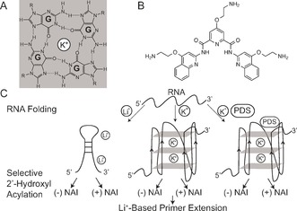
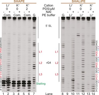
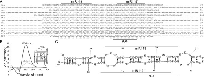
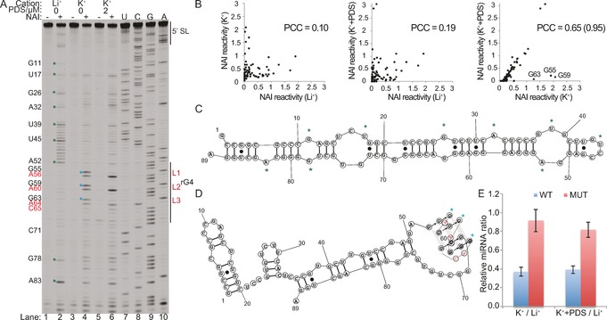
RNA G-quadruplex (rG4) structures are of fundamental importance to biology. A novel approach is introduced to detect and structurally map rG4s at single-nucleotide resolution in RNAs. The approach, denoted SHALiPE, couples selective 2'-hydroxyl acylation with lithium ion-based primer extension, and identifies characteristic structural fingerprints for rG4 mapping. We apply SHALiPE to interrogate the human precursor microRNA 149, and reveal the formation of an rG4 structure in this non-coding RNA. Additional analyses support the SHALiPE results and uncover that this rG4 has a parallel topology, is thermally stable, and is conserved in mammals. An in vitro Dicer assay shows that this rG4 inhibits Dicer processing, supporting the potential role of rG4 structures in microRNA maturation and post-transcriptional regulation of mRNAs.

Keywords: Dicer processing; G-quadruplexes; RNA structure; precursor miRNA; structure probing.


Figures

Figure 1
Figure 1
Structure probing of rG4s using SHALiPE. Chemical structure of A) a G‐quartet and B) pyridostatin (PDS). C) Overview of SHALiPE. RNA is folded under Li+, K+, or K++PDS conditions, followed by selective 2′‐hydroxyl acylation (SHA) using NAI. Li+‐based primer extension (LiPE) is conducted to analyze the NAI‐modified RNA. Controls are performed with no NAI.
Figure 2
Figure 2
SHALiPE development and analysis. TERRA RNA was probed with NAI under Li+, K+, and K++PDS conditions, followed by primer extension (PE) using either the SSIII commercial (com.) K+‐containing PE buffer14 (left gel) or home‐made Li+‐containing PE buffer (right gel). On the left, intense stalling was observed (lanes 1–6), caused by formation of TERRA rG4 (stabilized by K+ in the commercial PE buffer) in the PE step. On the right, stalling was reduced using the Li+‐based PE buffer. Lanes 9–14 show (−) and (+) NAI signals under Li+, K+, and K++PDS conditions. TERRA rG4 was unstructured in Li+ conditions. Under physiological K+ conditions, TERRA rG4 displayed distinct NAI profiles (low NAI signals in several Gs involved in rG4; purple asterisks, see lanes 10 and 12). Blue asterisks indicate Gs that were protected upon PDS addition (lanes 12 and 14). The loops (L1, L2, and L3) and loop nucleotides (UUA) are shown in red. Lanes 7–8 show sequencing of G. 5′ SL, 5′ stem loop (Supporting Information, Figure S1).
Figure 3
Figure 3
Computational analysis and biophysical characterization of a putative rG4 in human pre‐miRNA 149. A) Comparative sequence analysis of pre‐miRNA 149. The miR149, miR149*, and putative rG4 are highlighted. Conserved nucleotides are marked by asterisks and the length of RNAs are shown. The name of the species, presented here with three‐letter codes, can be found in the Supporting Information, Table S3. B) CD spectrum of wildtype rG4 sequence under K+ condition shows a characteristic signature that indicates formation of a parallel topology rG4 structure. Mutant shows no such signature. The Y axis is molar ellipticity per nucleotide (Δϵ). C) The secondary structure model for the human pre‐miRNA 149 is derived using the sequences in (A) and TurboFold.19
Figure 4
Figure 4
SHALiPE and Dicer cleavage assays on human pre‐miRNA 149 reveal an rG4 that inhibits in vitro Dicer processing. A) SHALiPE on pre‐miRNA 149 under Li+, K+, and K++PDS conditions (lanes 1–6). Lanes 7–10 show sequencing of U, C, G, and A respectively. The rG4 loops are in red. G that are protected with the addition of PDS (G55, G59, G63) are marked with blue asterisks. B) The PCC of the normalized NAI reactivity for K+ versus Li+ (left), K++PDS versus Li+ (middle), and K++PDS versus K+ (right). The PCC increases from 0.65 to 0.95 by removing G55, G59 and G63 (right). C) The hairpin conformation of pre‐miRNA 149. Some nucleotides are marked with green asterisks. D) The rG4‐containing conformation of pre‐miRNA 149. G55, G59, and G63 are marked with blue asterisks. E) Results of in vitro Dicer assay on pre‐miRNA 149 wildtype (WT) and mutant (MUT). Relative miRNA ratio (K+/Li+ or K++PDS/Li+) are reported. Values are from 3 biological replicates and error bars depict standard deviations. The constructs for the Dicer assay do not contain the 5′ and 3′SL (Supporting Information, Table S1).

Similar articles

Cited by

References

    1. None
    1. Millevoi S., Moine H., Vagner S., WIREs RNA 2012, 3, 495–507; - PubMed
    1. Simone R., Fratta P., Neidle S., Parkinson G. N., Isaacs A. M., FEBS Lett. 2015, 589, 1653–1668. - PubMed
    1. None
    1. Biffi G., Di Antonio M., Tannahill D., Balasubramanian S., Nat. Chem. 2014, 6, 75–80; - PMC - PubMed

Publication types

LinkOut - more resources