

Review
doi: 10.1056/NEJMra1511117.
Calcium Pyrophosphate Deposition Disease
Affiliations
- PMID: 27355536
- PMCID: PMC6240444
- DOI: 10.1056/NEJMra1511117
Item in Clipboard
Review
Calcium Pyrophosphate Deposition Disease
N Engl J Med.
.
No abstract available
Conflict of interest statement
No potential conflict of interest relevant to this article was reported.
Figures
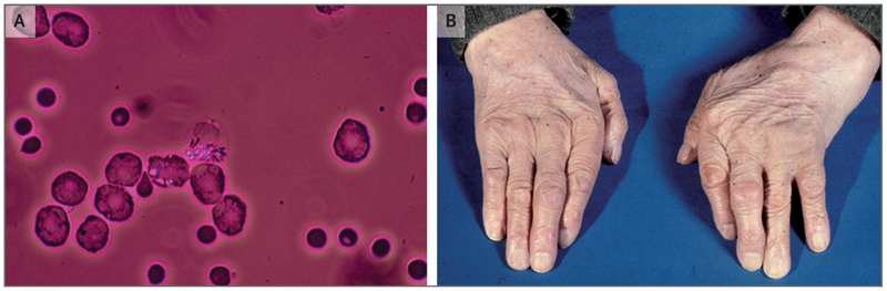
Rhomboid, birefringent calcium pyrophosphate (CPP) crystals are seen under polarizing light microscopy in this sample of synovial fluid that was obtained from a patient with acute CPP crystal arthritis of the wrist (Panel A). The hands of an elderly patient with CPPD disease show swelling in the left wrist and the third proximal interphalangeal joint of the left hand (Panel B).
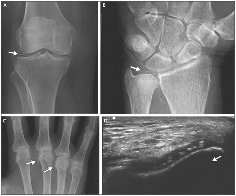
Panel A shows a radiograph of a knee with meniscal chondrocalcinosis (arrow). Panel B shows a radiograph of a wrist with chondrocalcinosis of the triangular cartilage (arrow). Panel C shows a radiograph of a hand with hooklike osteophytes (arrows). Panel D shows an ultrasonographic image of a right knee, which was obtained with the transducer in the anatomical axial plane, with the knee flexed 90 degrees. The probe was pointed at the femoral cartilage on the “V” of the patellar groove. Chondrocalcinosis is seen in the substance of the cartilage; the arrow indicates the direction of the probe.
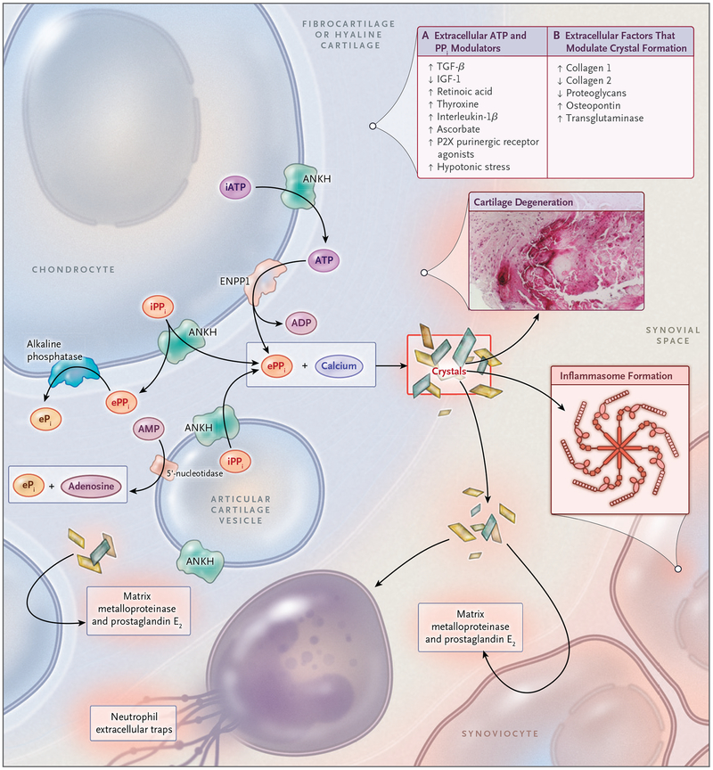
The formation of CPP crystals occurs in the articular cartilage pericellular matrix and is facilitated by extracellular vesicles known as articular cartilage vesicles. Pyrophosphate (PPi) is generated from extracellular ATP and forms complexes with calcium to create CPP crystals. Panel A of the box, upper right, lists the factors that are known to modulate levels of extracellular ATP and PPi, and Panel B the extracellular matrix factors that regulate the formation of CPP crystals. P2X indicates one class of purinergic receptors. CPP crystals induce inflammation in the synovial space but also have adverse biomechanical consequences and direct catabolic effects on joint tissues owing to the production of prostaglandin E2 and matrix metalloproteinases. These factors ultimately produce cartilage degeneration, as shown by the CPP crystal deposit in cartilage in situ (inset). ANKH denotes human homologue of the protein product of the progressive ankylosis gene, ENPP1 ectonucleotide pyrophosphatase 1, ePi extracellular phosphate ion, ePPi extracellular PPi, IGF-1 insulin-like growth factor 1, iPPi intracellular PPi, and TGF-β transforming growth factor β.
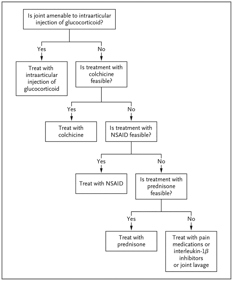
A treatment is considered to be feasible if it is not associated with unacceptable side effects. NSAID denotes nonsteroidal antiinflammatory drug.
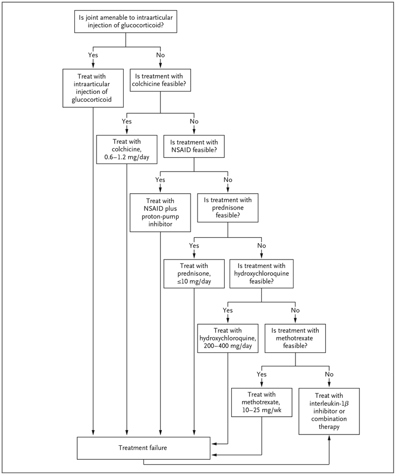
Combination therapy may include various combinations of colchicine, prednisone, methotrexate, and hydroxychloroquine.
References
-
- McCarty D Jr, Kohn NN, Faires JS. The significance of calcium pyrophosphate crystals in the synovial fluid of arthritic patients: the “pseudogut syndrome.” 1. Clinical aspects. Ann Intern Med 1962; 56: 711–37. - PubMed
-
- Zhang W, Doherty M, Bardin T, et al. European League Against Rheumatism recommendations for calcium pyrophosphate deposition. I. terminology and diagnosis. Ann Rheum Dis 2011; 70: 563–70. - PubMed
-
- Masuda I, Ishikawa K. Clinical features of pseudogout attack: a survey of 50 cases. Clin Orthop Relat Res 1988; 229: 173–81. - PubMed
-
- McCarty D Jr. Diagnostic mimicry in arthritis: patterns of joint involvement associated with calcium pyrophosphate dihydrate crystal deposits. Bull Rheum Dis 1975; 25: 804–9.
Publication types
MeSH terms
Substances
Grants and funding
LinkOut - more resources
Full Text Sources
Other Literature Sources