Review
. 2016 Jun 27;5(3):29.
doi: 10.3390/cells5030029.

Epithelial Intermediate Filaments: Guardians against Microbial Infection?

Affiliations
Review

Epithelial Intermediate Filaments: Guardians against Microbial Infection?

Florian Geisler et al. Cells. .

Abstract

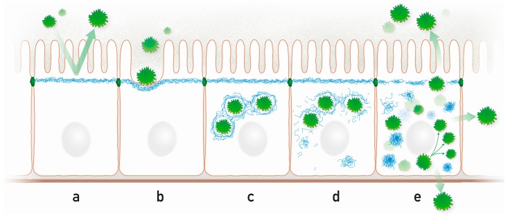
Intermediate filaments are abundant cytoskeletal components of epithelial tissues. They have been implicated in overall stress protection. A hitherto poorly investigated area of research is the function of intermediate filaments as a barrier to microbial infection. This review summarizes the accumulating knowledge about this interaction. It first emphasizes the unique spatial organization of the keratin intermediate filament cytoskeleton in different epithelial tissues to protect the organism against microbial insults. We then present examples of direct interaction between viral, bacterial, and parasitic proteins and the intermediate filament system and describe how this affects the microbe-host interaction by modulating the epithelial cytoskeleton, the progression of infection, and host response. These observations not only provide novel insights into the dynamics and function of intermediate filaments but also indicate future avenues to combat microbial infection.

Keywords: Caenorhabditis elegans; bacterium; barrier; epithelium; keratin; parasite; pathogen; virus.


Conflict of interest statement

The authors declare no conflict of interest.

Figures

Figure 1
Figure 1
IFs form complex three-dimensional networks with cell type specific subcellular arrangements providing barrier function in simple and stratified epithelia. (A) Keratin IFs (blue) are subapically enriched in a dense filamentous network in the simple epithelium of the intestine. They localize just below the microvillar brush border that protrudes into the nutrient-filled intestinal lumen. The cylindrical epithelial cells are connected by junctional complexes, which encompass keratin-anchoring desmosomes (green), and rest all on a basal lamina; (B) The keratin IFs (blue) of the stratified epithelium of the cornea form dense 3D-networks that traverse the entire cytoplasm and are attached to desmosomes (green) at cell-cell contact sites. Keratin fragments with antibacterial activity are released into the tear fluid. The keratin cytoskeleton of the basal cells is anchored to hemidesmosomes (red), which attach to the underlying extracellular matrix of the basement membrane; (C) The keratin IF cytoskeleton of the epidermis, which is the prototype of a multilayered cornified epithelium, increases in density in the flattened suprabasal cell layers and becomes compacted as part of the cornified envelope of the dead cells in the uppermost stratum corneum which are continuously shed from the epithelium. While desmosomes (green) are present in all cell layers, hemidesmosomes (red) are restricted to the cuboidal basal cell layer.
Figure 2
Figure 2
Highly schematic representation of processes that may occur during microbe-intermediate filament interaction in a simple epithelium. (a) The subapically enriched cytoplasmic IF system acts as an intracellular protective barrier; (b) IFs form together with the actin cytoskeleton pedestals for attached microbes; (c) Intracellular microbes are encaged by IFs; (d) Microbes disrupt the IF cage through kinase activities, which modify IF polypeptides and initiate the formation of cytoplasmic IF aggregates; (e) Released microbes proliferate and spread to neighboring cells and to the environment disrupting the protective apical IF network. Prominent cytoplasmic aggregates containing hyperphosphorylated IFs appear.

Similar articles

Cited by

References

    1. Kim S., Coulombe P.A. Intermediate filament scaffolds fulfill mechanical, organizational, and signaling functions in the cytoplasm. Genes Dev. 2007;21:1581–1597. doi: 10.1101/gad.1552107. - DOI - PubMed
    1. Magin T.M., Vijayaraj P., Leube R.E. Structural and regulatory functions of keratins. Exp. Cell Res. 2007;313:2021–2032. doi: 10.1016/j.yexcr.2007.03.005. - DOI - PubMed
    1. Toivola D.M., Strnad P., Habtezion A., Omary M.B. Intermediate filaments take the heat as stress proteins. Trends Cell Biol. 2010;20:79–91. doi: 10.1016/j.tcb.2009.11.004. - DOI - PMC - PubMed
    1. Margiotta A., Bucci C. Role of intermediate filaments in vesicular traffic. Cells. 2016;5:20. doi: 10.3390/cells5020020. - DOI - PMC - PubMed
    1. Herrmann H., Bar H., Kreplak L., Strelkov S.V., Aebi U. Intermediate filaments: From cell architecture to nanomechanics. Nat. Rev. Mol. Cell Biol. 2007;8:562–573. doi: 10.1038/nrm2197. - DOI - PubMed

LinkOut - more resources